Please use this identifier to cite or link to this item:
https://hdl.handle.net/2445/222923| Title: | Inner structure of the tongue |
| Other Titles: | Organització interna de la llengua |
| Author: | Pérez Clausell, Jeús Universitat de Barcelona. Departament de Biologia Cel·lular, Fisiologia i Immunologia |
| Keywords: | Llengua (Anatomia) Aparell digestiu Epiteli Nervis Histologia Vasos sanguinis Múscul estriat |
| Issue Date: | 2025 |
| Citation: | Cite as: Pérez-Clausell J (2025). Inner structure of the tongue. Universitat de Barcelona. Departament de Biologia Cel·lular, Fisiologia i Immunologia. Dipòsit Digital UB [consulted on <<date>>] https://hdl.handle.net/2445/222923 |
| Abstract: | [ENG] A collection of micrographs illustrating the organisation of the tongue in mice and rats. The stratified epithelium, connective tissue layers, muscular bundles, blood vessels, and nerves are shown in paraffin sections stained with hæmatoxylin-eosin or van Gieson trichrome. Semithin sections 2 µm thick were stained with toluidine blue. This collection complements the micrographs published by Dr Elena Sagristà i Mateo https://hdl.handle.net/2445/60015 — See also: https://diposit.ub.edu/dspace/browse?type=author&value=Sagrist%C3%A0+i+Mateo%2C+Elena [CAT] Col·lecció de micrografies de l'organització de la llengua del ratolí i de la rata. Es mostra l’epiteli estratificat, el teixit conjuntiu, els feixos musculars, vasos sanguinis i nervis en talls de parafina tenyits amb hematoxilina-eosina, el tricròmic de van Gieson o talls semifins de resina epoxi tenyits amb blau de toluïdina. Aquesta col·lecció complementa les micrografies publicades per la Dra. Elena Sagristà i Mateo https://hdl.handle.net/2445/60015 — I també: https://diposit.ub.edu/dspace/browse?type=author&value=Sagrist%C3%A0+i+Mateo%2C+Elena |
| URI: | https://hdl.handle.net/2445/222923 |
| Appears in Collections: | OMADO (Objectes i MAterials DOcents) |
Files in This Item:
| File | Description | Size | Format | |
|---|---|---|---|---|
| P_O_tongue_HEosin_2x_b41_b44_collage_fused_LETTERS.jpg | Transverse section of the body of a mouse tongue. The epithelium (ep) is thicker on the dorsal surface (top in the figure) and lateral surfaces, forming cone-shaped protrusions, the filiform papillae (asterisks). The epithelium on the ventral surface (bottom in the figure) is thinner and lacks papillae. Beneath the epithelium lies the lamina propria (lp), a connective tissue layer. This level of the tongue is close to its insertion into the floor of the oral cavity, and the ventral lamina propria (lpˬv) is thick and lightly stained, containing sections of the deep lingual vein (v, filled with erythrocytes). Beneath the lamina propria, a large mass of striated muscles is observed in transverse section: the superior longitudinal muscles (sl) dorsally, and the inferior longitudinal muscles (il) ventrally. Other muscular bundles are seen in longitudinal section: the massive transverse muscles (t) and the vertical muscles (ve), in thin bundles (nomenclature as in McClung & Goldberg, 2000). A portion of the genioglossus muscle (ge) which attaches the tongue to the floor of the oral cavity, is also visible. Paraffin section, hæmatoxylin-eosin stain. a, adipose tissue; ar, arteries. | 1 MB | JPEG |  View/Open |
| P_O_tongue_HEosin_2x_b41_b44_collage_fused.jpg | Transverse section of the body of a mouse tongue. No labelling. | 966.67 kB | JPEG | 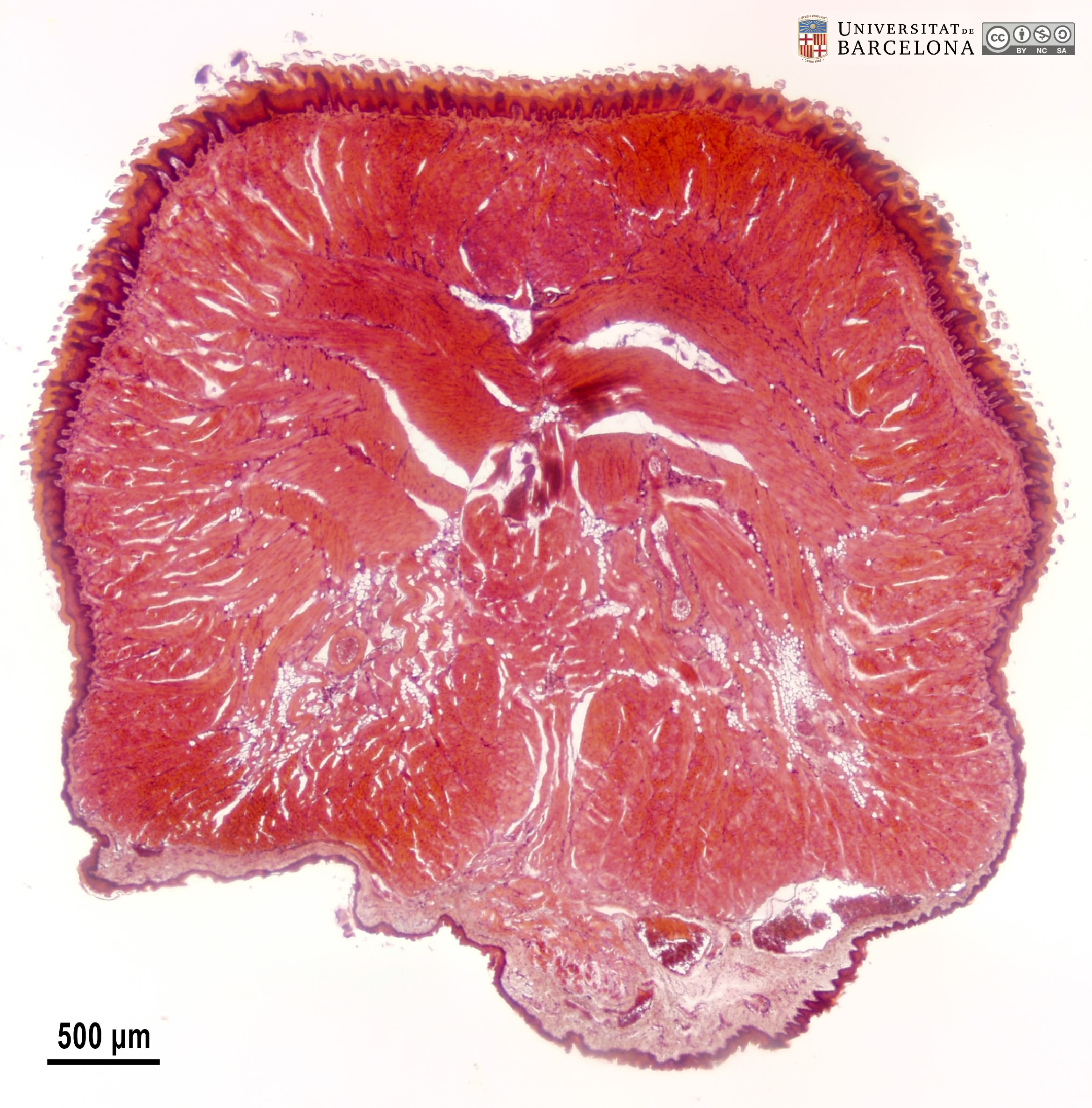 View/Open |
| P_O_tongue_HEosin_vGieson_4x_combinat_2_LETTERS.jpg | Collage of two sections of the tip (apex) of the mouse tongue stained with hæmatoxylin-eosin (left) and van Gieson trichrome (right). The epithelium (ep) is distinctly stained in both micrographs, and forms cone-shaped elevations, the filiform papillae (asterisks). On the right (van Gieson), beneath the epithelium, the collagen fibres in the lamina propria (lp) are stained dark pink or red with acid fuchsin. In the left micrograph (hæmatoxylin-eosin), the lamina propria (lp) appears light pink and is difficult to distinguish from the underlying muscle fibres. In contrast, in the van Gieson trichrome (right) the fibres of the superior longitudinal muscles (sl), transverse muscles (t), and inferior longitudinal muscles (il) are stained brown and can be distinguished from the connective tissue (pink) in the lamina propria and that between the muscular bundles. Paraffin section, hæmatoxylin-eosin (left) and van Gieson (right). ar, artery; n, nerve branches; v, veins. | 1.68 MB | JPEG | 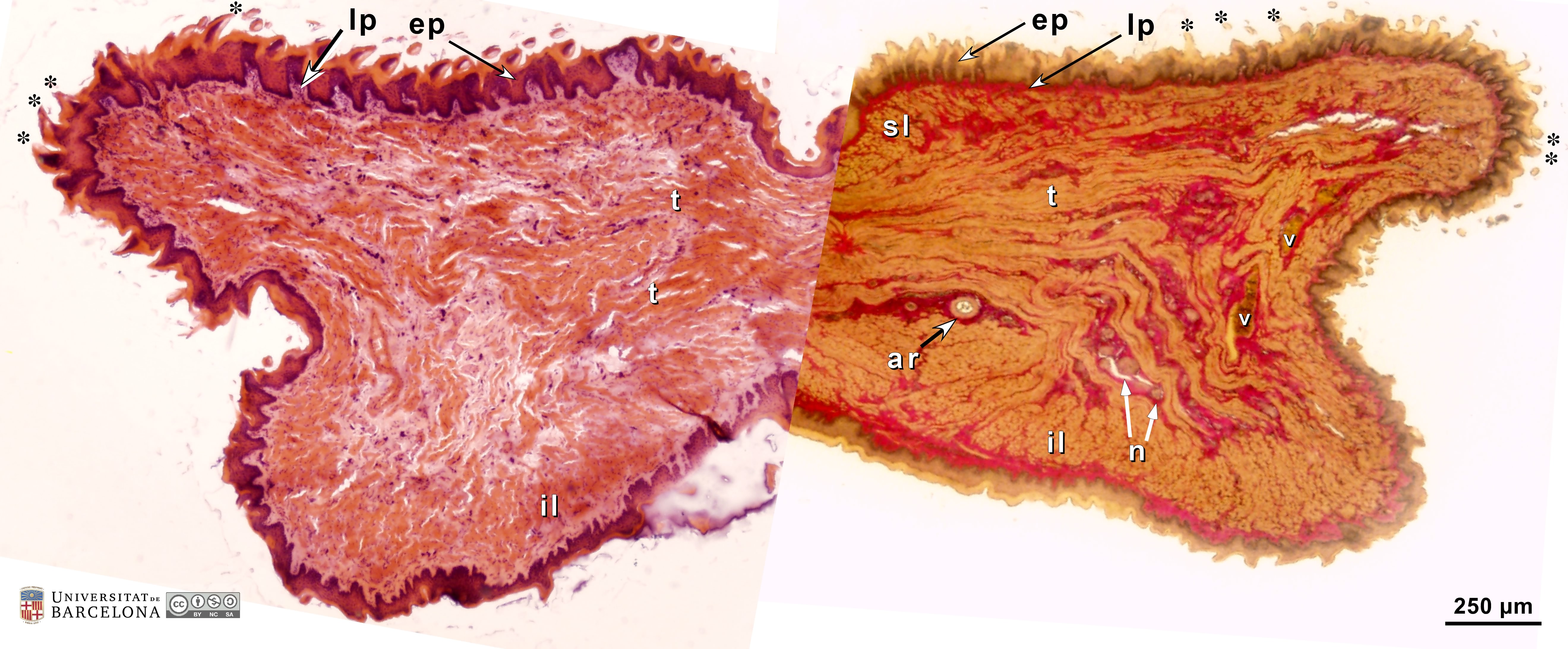 View/Open |
| P_O_tongue_HEosin_vGieson_4x_combinat.jpg | Collage of two sections of the tip (apex) of the mouse tongue stained with hæmatoxylin-eosin (left) and van Gieson trichrome (right). No labelling. | 1.63 MB | JPEG |  View/Open |
| P_O_tongue_HEosin_4x_b50_LETTERS.jpg | Dorsal surface of the mouse tongue in transverse section. The epithelium (ep) is unevenly thick, with conical protrusions, the filiform papillae (asterisks). In addition to the living epithelial cells stained purple, a stratum corneum (cor) of dead cells accumulates on the surface of the mouse epithelium (light orange). Immediately below the epithelium there is a thin layer of connective tissue – the lamina propria (lp) – stained pale pink, which projects into the epithelium contributing to the papillae (white stars). Deeper in the tongue there are bundles of skeletal (striated) muscles (st): massive bundles of the superior longitudinal (sl) and transverse (t) muscles and thin strips of vertical muscles (ve). Paraffin section, hæmatoxylin-eosin. ar, artery. | 997.07 kB | JPEG | 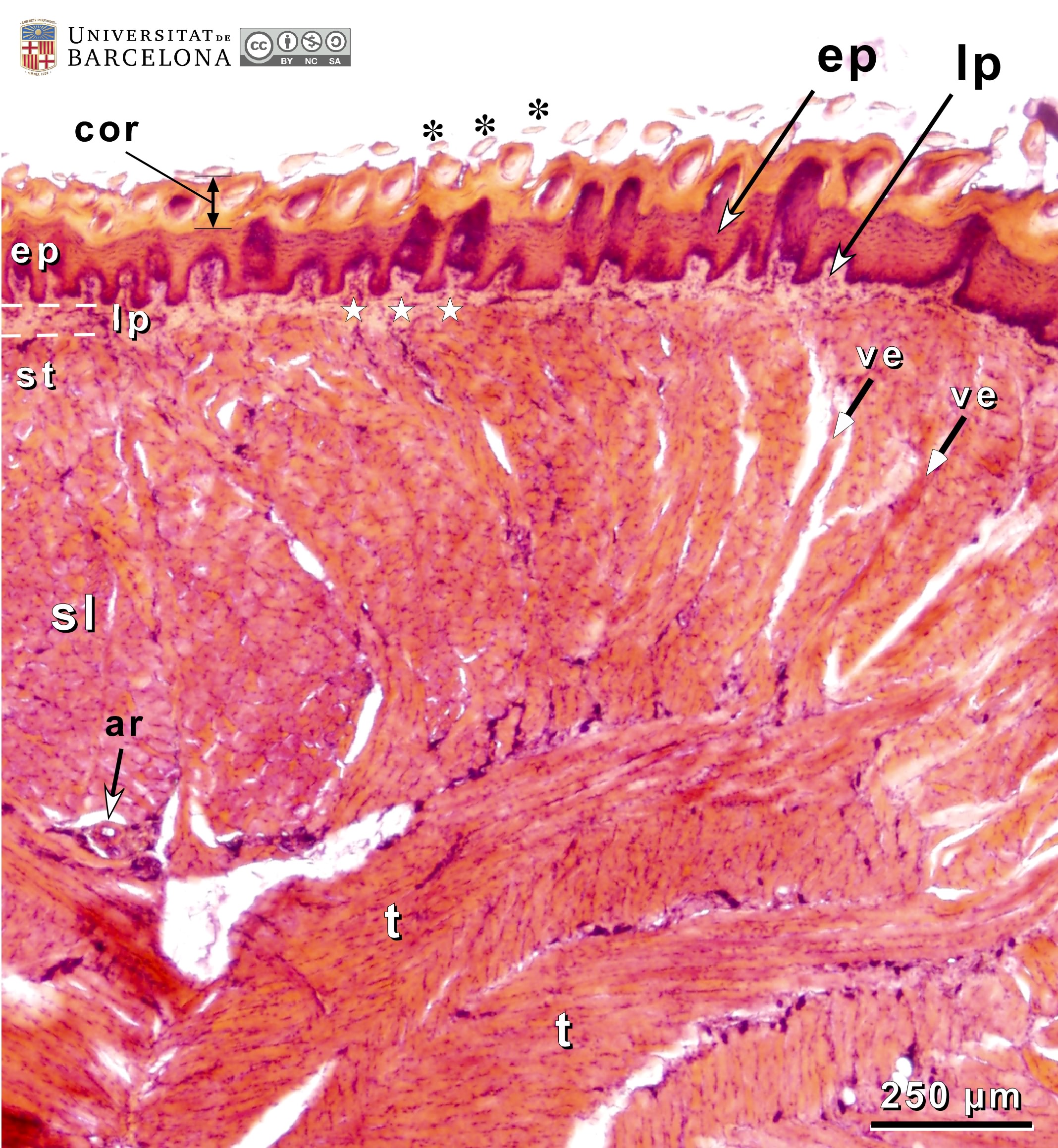 View/Open |
| P_O_tongue_HEosin_4x_b50.jpg | Dorsal surface of the mouse tongue in transverse section. No labelling. | 959.79 kB | JPEG | 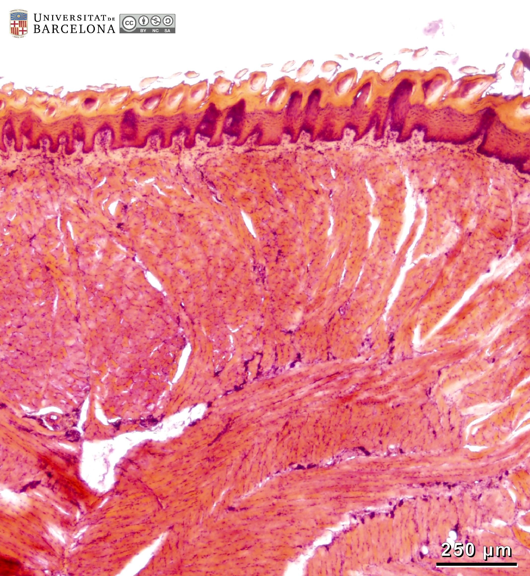 View/Open |
| P_O_tongue_HEosin_20x_28_LETTERS.jpg | Dorsal surface of the mouse tongue. A stratified squamous epithelium (ep) is observed. It contains a stratum basale (bas) or germinative layer with darkly stained, purple nuclei, a stratum spinosum (sp) although cells do not look spiny here, a stratum granulosum (gra) in which small, purple granules can be seen, and a thick stratum corneum (cor) of aggregated dead cells stained orange. The lamina propria (lp) forms a thin layer of connective tissue lightly stained, with protrusions of this connective tissue (empty stars) towards the overlying epithelium contributing to the emergence of the papillae (asterisks). Beneath the lamina propria, skeletal (striated) muscle fibres are seen in transverse sections (striae not visible as a result). Paraffin section, hæmatoxylin-eosin stain. | 844.97 kB | JPEG | 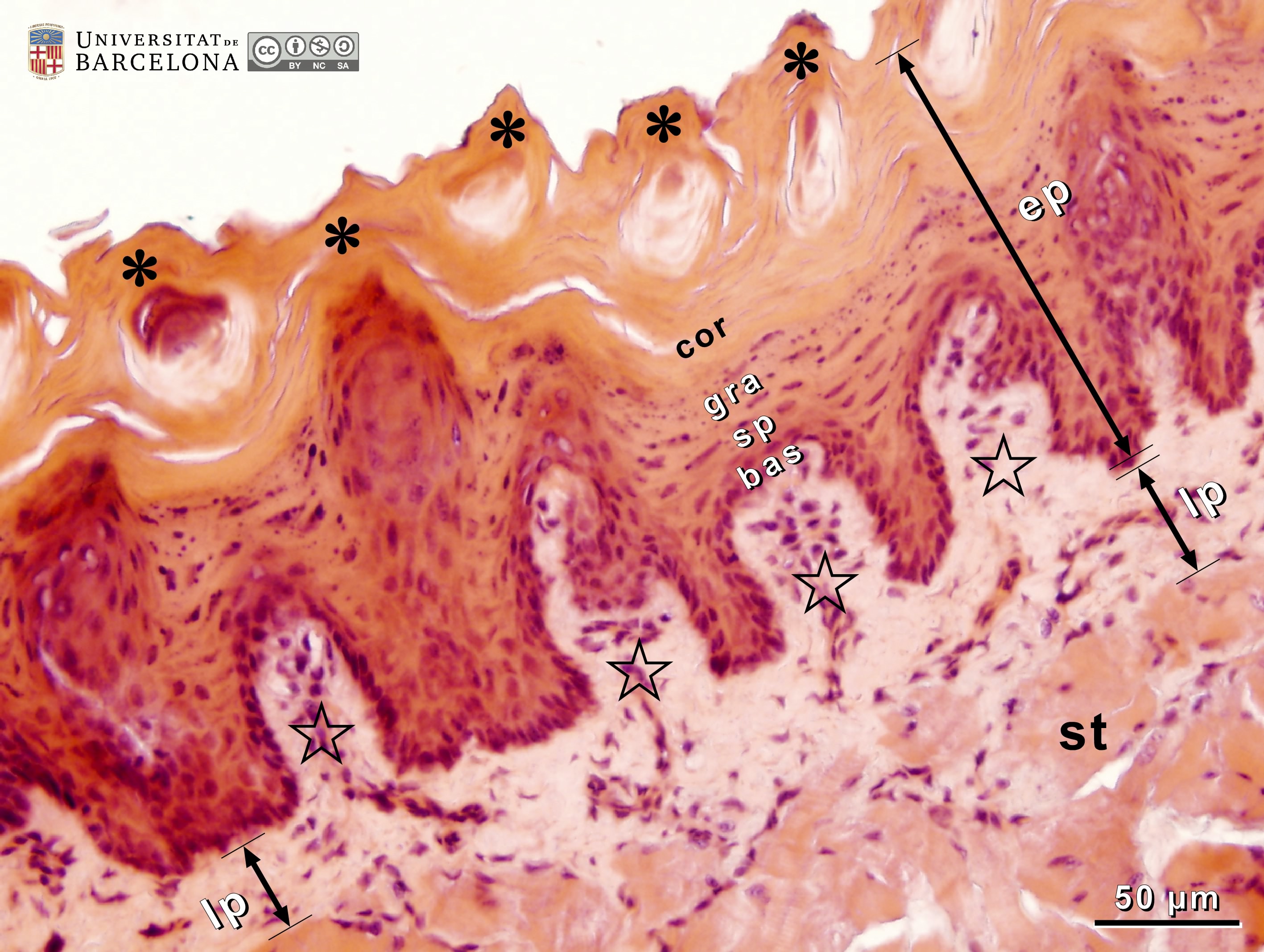 View/Open |
| P_O_tongue_HEosin_20x_28.jpg | Dorsal surface of the mouse tongue. No labelling. | 785.8 kB | JPEG | 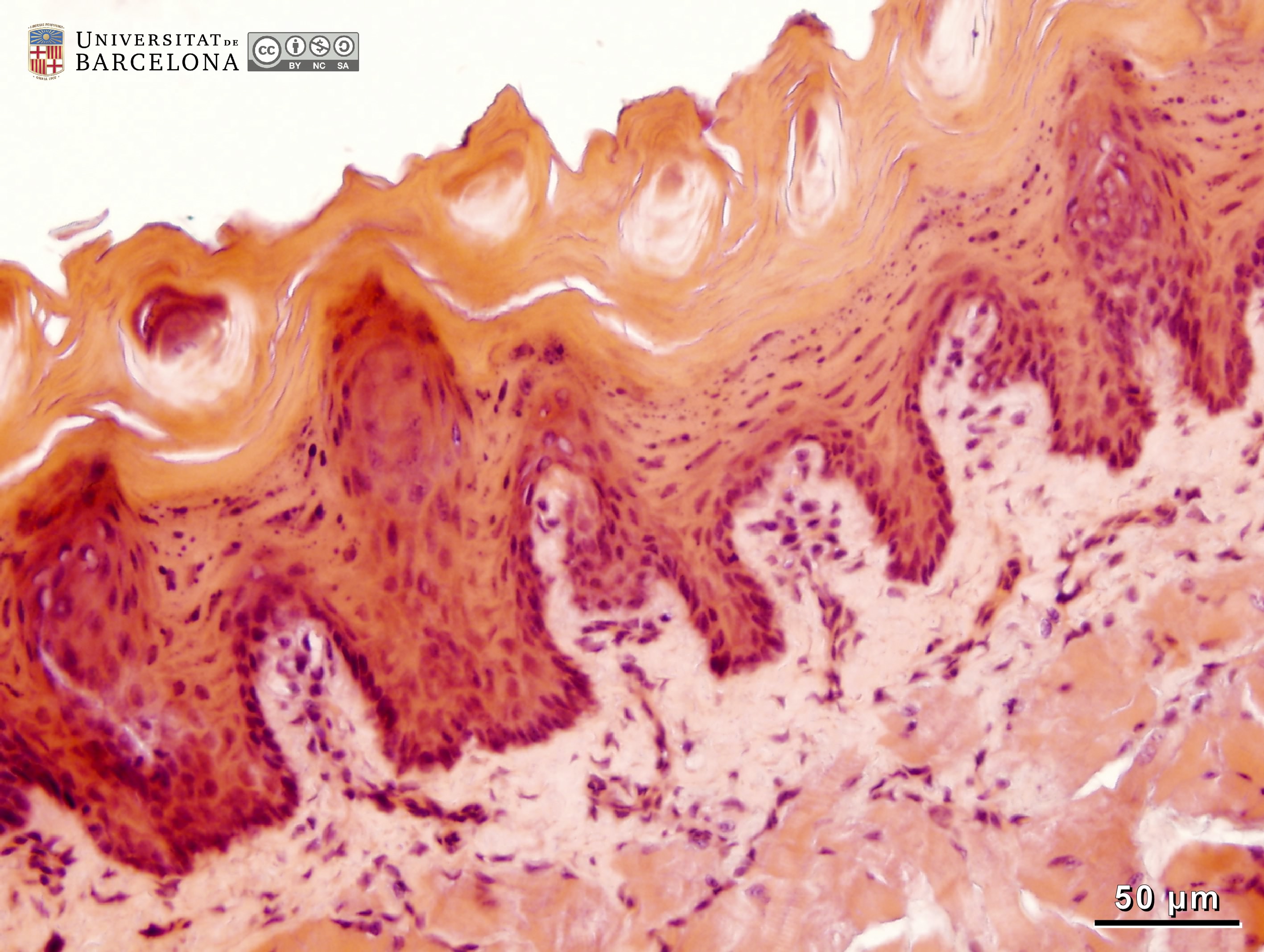 View/Open |
| P_O_tongue_HEosin_50x_49_51_collage_LETTERS.jpg | Dorsal surface of the mouse tongue at high magnification (collage of micrographs allowing detailed zoom). Both the epithelium (ep) and the lamina propria (lp) have an undulating appearance which generates the lingual papillae. The epithelium consists of the stratum basale (bas) with darkly stained, round cell nuclei, stratum spinosum with paler, elongated nuclei, stratum granulosum with paler nuclei and purple keratohyalin granules, and stratum corneum (cor) of anucleated, dead cells forming a multilayered, brightly stained zone. The conical projections of the epithelium form the filiform papillae (asterisks at the tip of two of them). In the lamina propria (lp), darkly stained cells are scattered in a lightly stained extracellular matrix. Upward expansions of the lamina propria into the overlying epithelium (empty stars) contribute to the papillary organisation. Deep to the lamina propria, bundles of skeletal muscles are weakly stained. When cut longitudinally, the striated appearance of the muscle fibres is faintly noticeable (empty arrowheads). Paraffin section, hæmatoxylin-eosin stain. | 1.06 MB | JPEG | 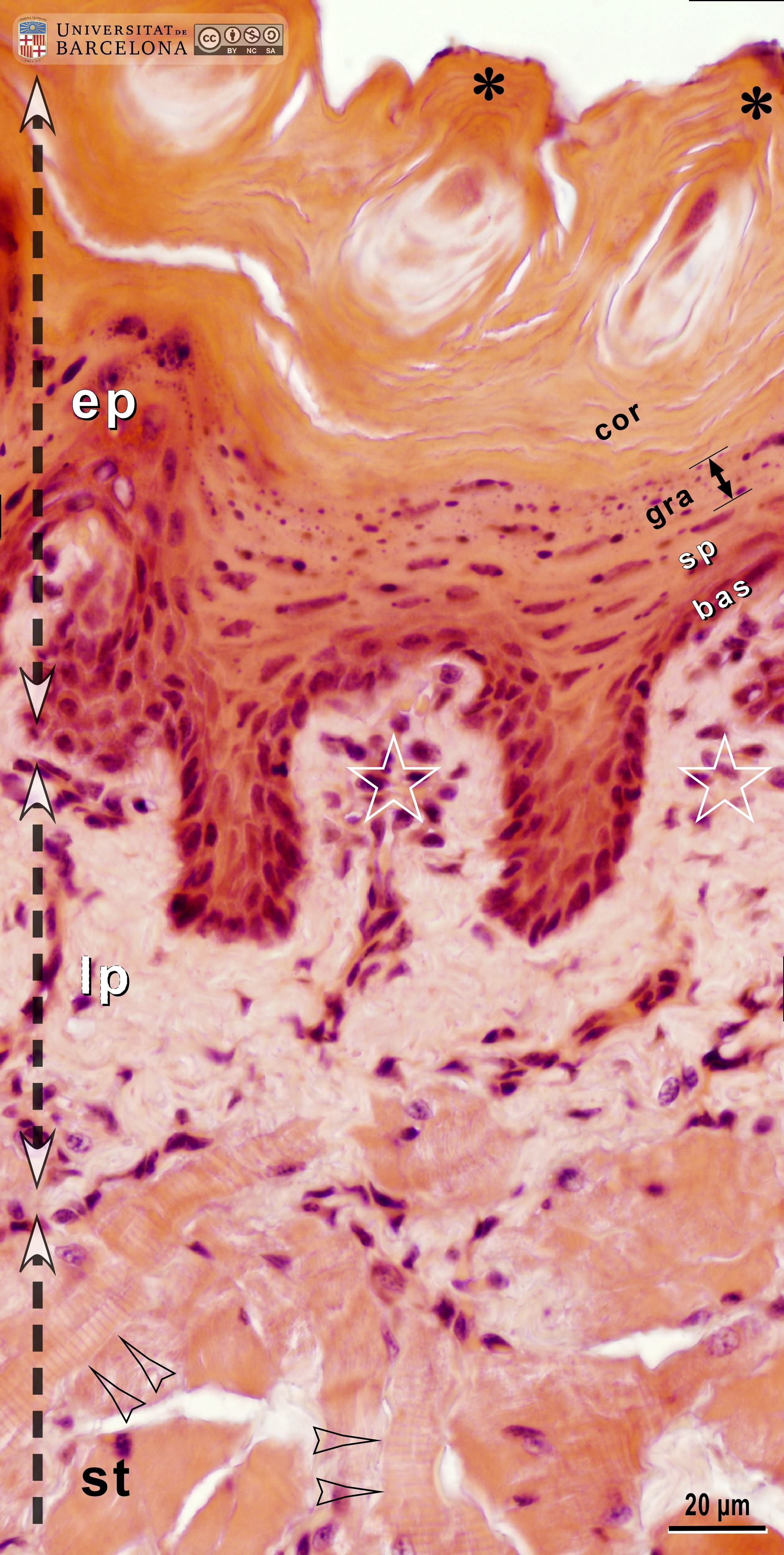 View/Open |
| P_O_tongue_HEosin_50x_49_51_collage.jpg | Dorsal surface of the mouse tongue at high magnification (collage of micrographs allowing detailed zoom). No labelling. | 1 MB | JPEG | 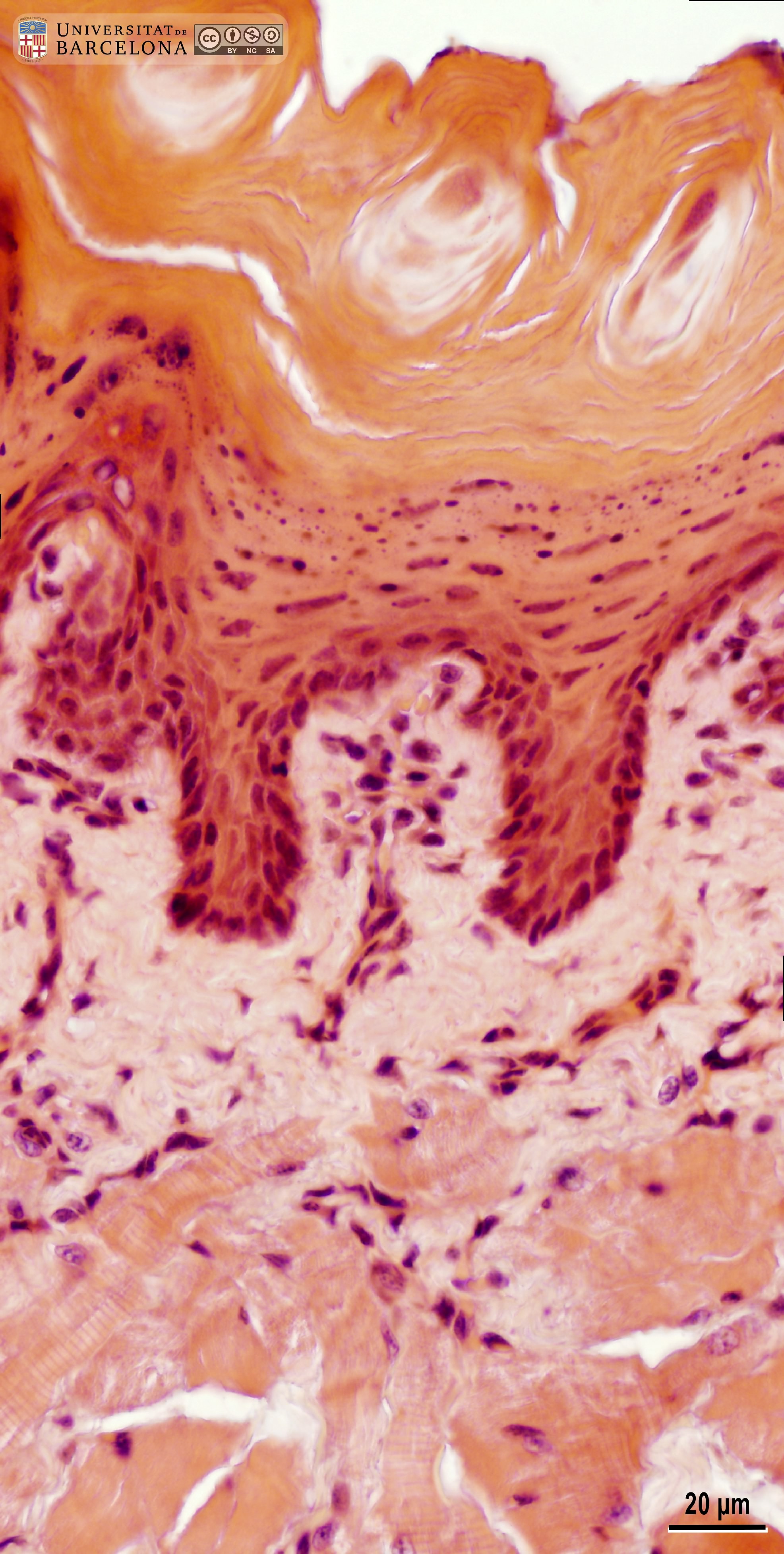 View/Open |
| P_O_tongue_HEosin_50x_40_42_collage_LETTERS.jpg | Ventral surface of the mouse tongue at high magnification (collage of micrographs allowing detailed zoom). The micrograph was flipped vertically to facilitate layer comparison. The epithelium is thinner than that on the dorsal surface, but the same layers can be recognised: a stratum basale (bas), with dark nuclei, an intermediate stratum spinosum (sp), with paler, elongated cell nuclei, stratum granulosum (gra), with deep purple keratohyalin granules, and a thin but distinct stratum corneum, of anucleated, dead cells. Immediately below the epithelium, a thick lamina propria (lp) is highly vascularised, and capillaries (c) and veins of various sizes (v), some of them with erythrocytes (er) inside, can be observed. Here, darkly stained cells are scattered in a lightly stained, pink matrix containing collagen fibres (black arrowheads). Beneath this, bundles of skeletal muscle fibres (st) are present, and their striae can be barely noticed in longitudinal sections (empty arrowhead). Paraffin section, hæmatoxylin-eosin stain. | 1.06 MB | JPEG | 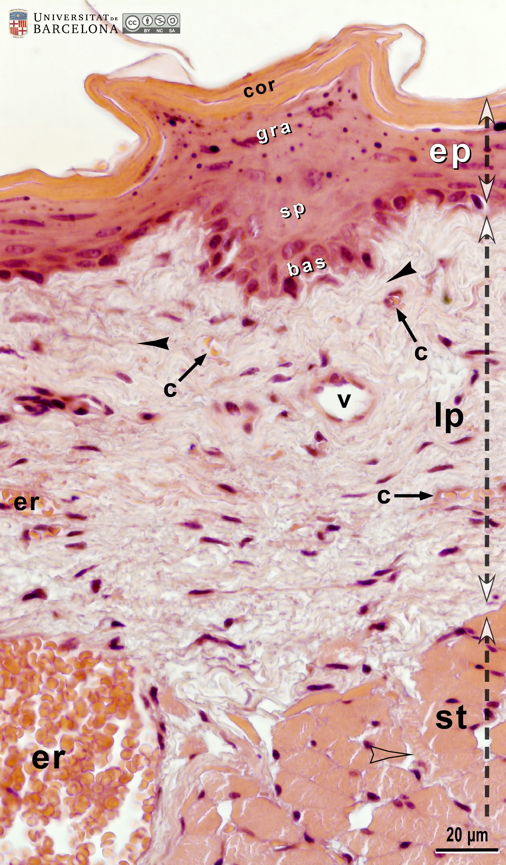 View/Open |
| P_O_tongue_HEosin_50x_40_42_collage.jpg | Ventral surface of the mouse tongue at high magnification (collage of micrographs allowing detailed zoom). No labelling. | 1.01 MB | JPEG |  View/Open |
| P_O_tongue_HEosin_50x_43_LETTERS.jpg | Ventral surface of the mouse tongue (micrograph flipped vertically for layer comparison). Papillae are not present here. This smooth surface consists of a stratified squamous epithelium (ep) including a stratum basale with darkly stained cell nuclei (N), an intermediate stratum spinosum (not labelled) with flattened, lighter nuclei, an overlying stratum granulosum (gra) with purple keratohyaline granules and a superficial stratum corneum (cor) composed of multiple layers of anucleated dead cells stained orange. The connective tissue layer – lamina propria (lp) – contains lightly stained collagen fibres (black arrowheads), scattered cells, and capillary vessels (c) enclosing some erythrocytes stained bright orange. Paraffin section, hæmatoxylin-eosin stain. er, erythrocytes within a large vein. | 770.21 kB | JPEG | 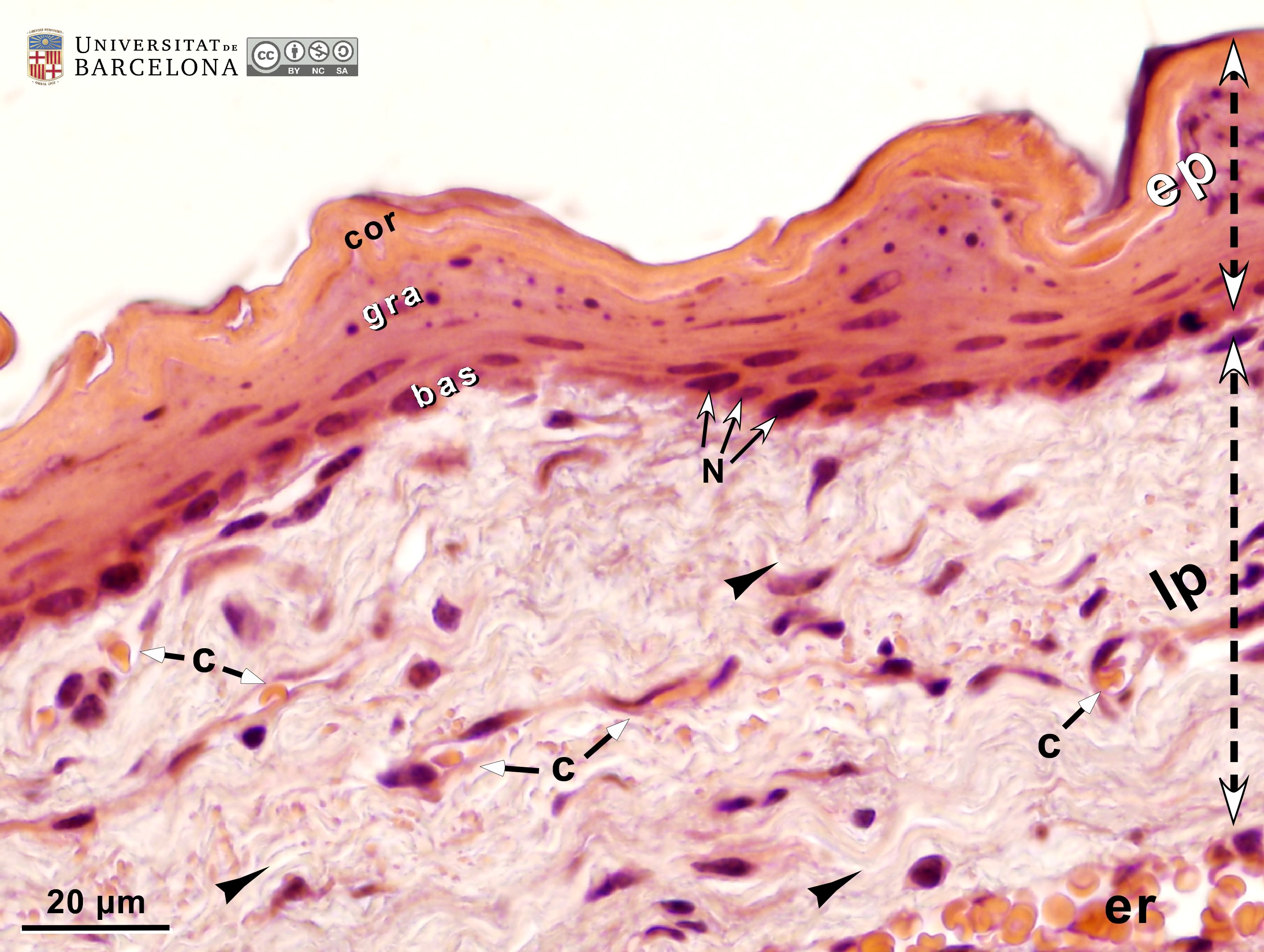 View/Open |
| P_O_tongue_HEosin_50x_43.jpg | Ventral surface of the mouse tongue (micrograph flipped vertically for layer comparison). No labelling. | 726.39 kB | JPEG |  View/Open |
| P_O_tongue_HEosin_10x_34_LETTERS.jpg | Vascularisation and innervation in the mouse tongue. Several nerve branches (n) and an artery (ar) can be seen in the connective tissue between the striated muscle fibre bundles (st). White adipocytes (a) are present in this interstitial connective tissue. Paraffin section, hæmatoxylin-eosin stain. ta, tunica adventitia; tm, tunica media. * For details on arteries, see figure → https://diposit.ub.edu/dspace/bitstream/2445/222162/1/P_O_tongue_HEosin_20x_35_LETTERS.jpg ← and others in → https://hdl.handle.net/2445/222162 ← | 915.94 kB | JPEG | 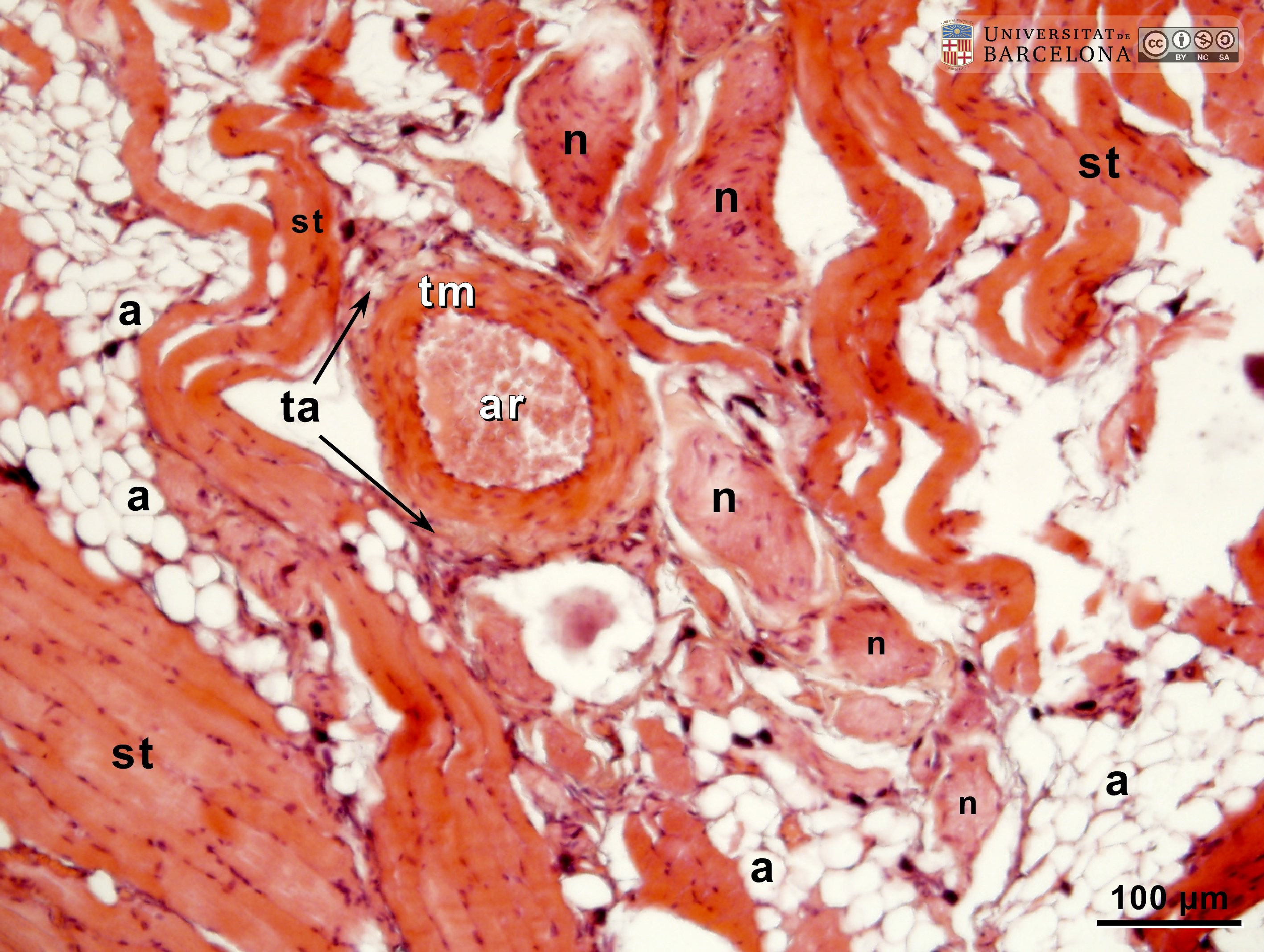 View/Open |
| P_O_tongue_HEosin_10x_34.jpg | Vascularisation and innervation in the mouse tongue. No labelling. | 892.37 kB | JPEG | 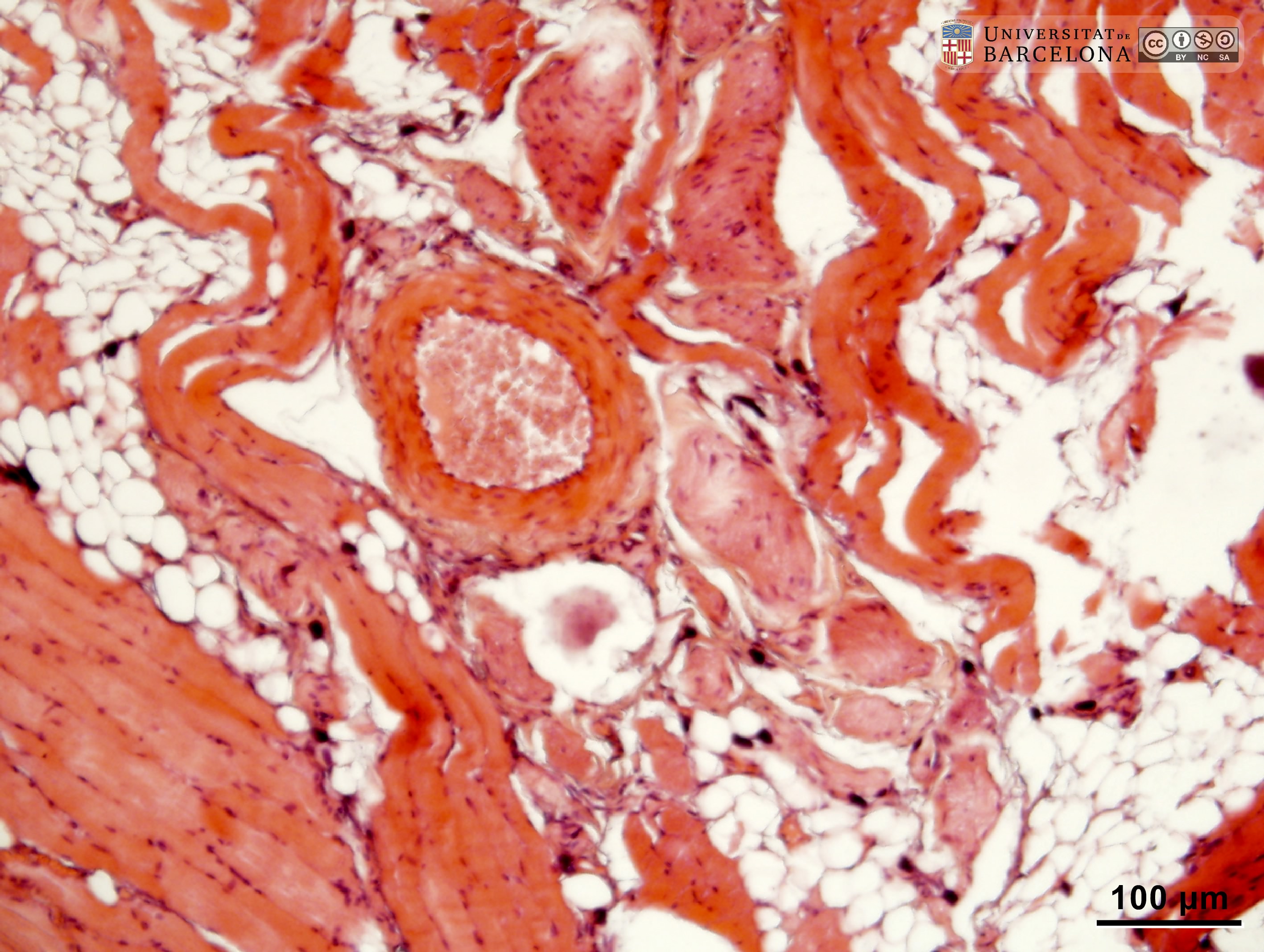 View/Open |
| P_O_tongue_HEosin_50x_45_LETTERS2.jpg | Nerve close to a skeletal muscle fibre in the mouse tongue. In this longitudinal section of the muscle fibre, cell nuclei are dark and located in the periphery of the tubular fibre (arrows) while the central region of the tube (st) shows a transverse striated pattern. The adjacent nerve (n) is surrounded by a layer of lightly stained collagen fibres (black arrowheads). Within the nerve, white, round profiles probably represent the myelinated axon sheaths (empty arrowheads). Paraffin section, hæmatoxylin-eosin stain. | 696.54 kB | JPEG | 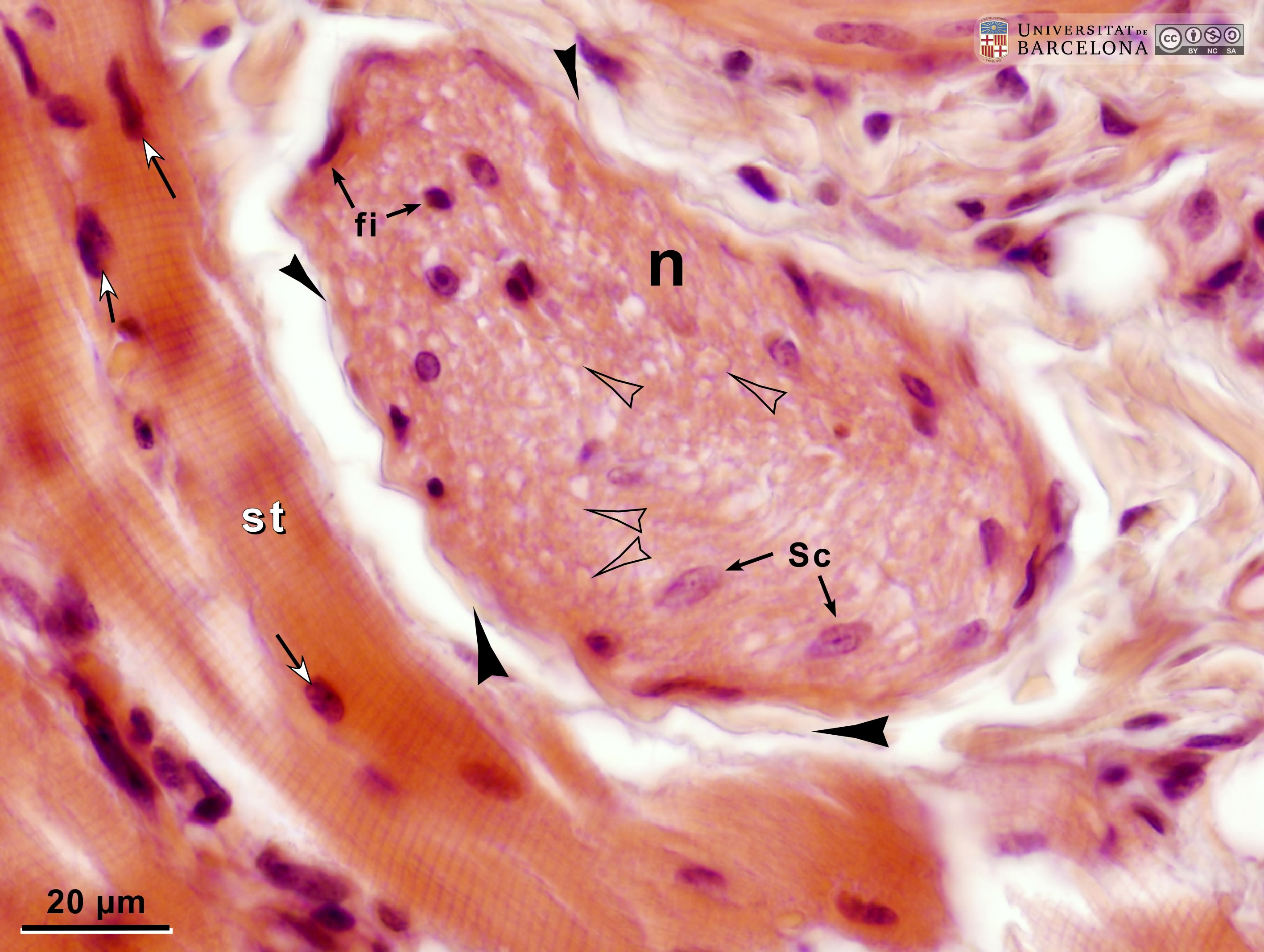 View/Open |
| P_O_tongue_HEosin_50x_45.jpg | Nerve close to a skeletal muscle fibre in the mouse tongue. No labelling. | 668.8 kB | JPEG | 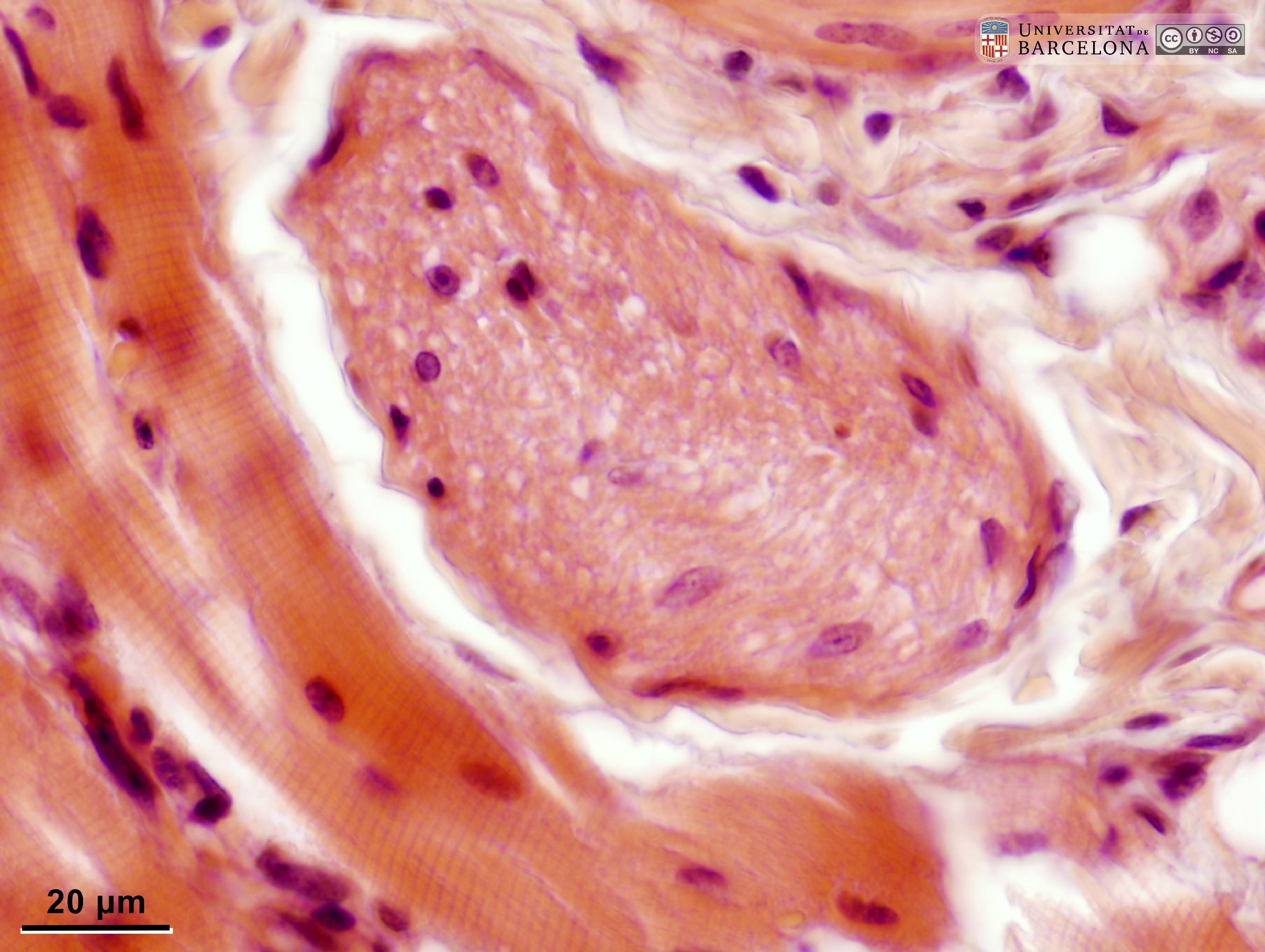 View/Open |
| P_O_tongue_HEosin_50x_38_LETTERS.jpg | Skeletal muscle in the mouse tongue. Fibres of the superior longitudinal muscle (sl) are seen in transverse section, and the cell nuclei are deep purple and located in the periphery of the fibre (arrows). Below, fibres of the transverse muscle (t) are cut longitudinally. Nuclei are elongated and located in the periphery of the fibres as above (arrows). In contrast to the transverse sections, a distinct striated pattern can be seen within the muscle fibres. Paraffin section, hæmatoxylin-eosin stain. | 775.89 kB | JPEG | 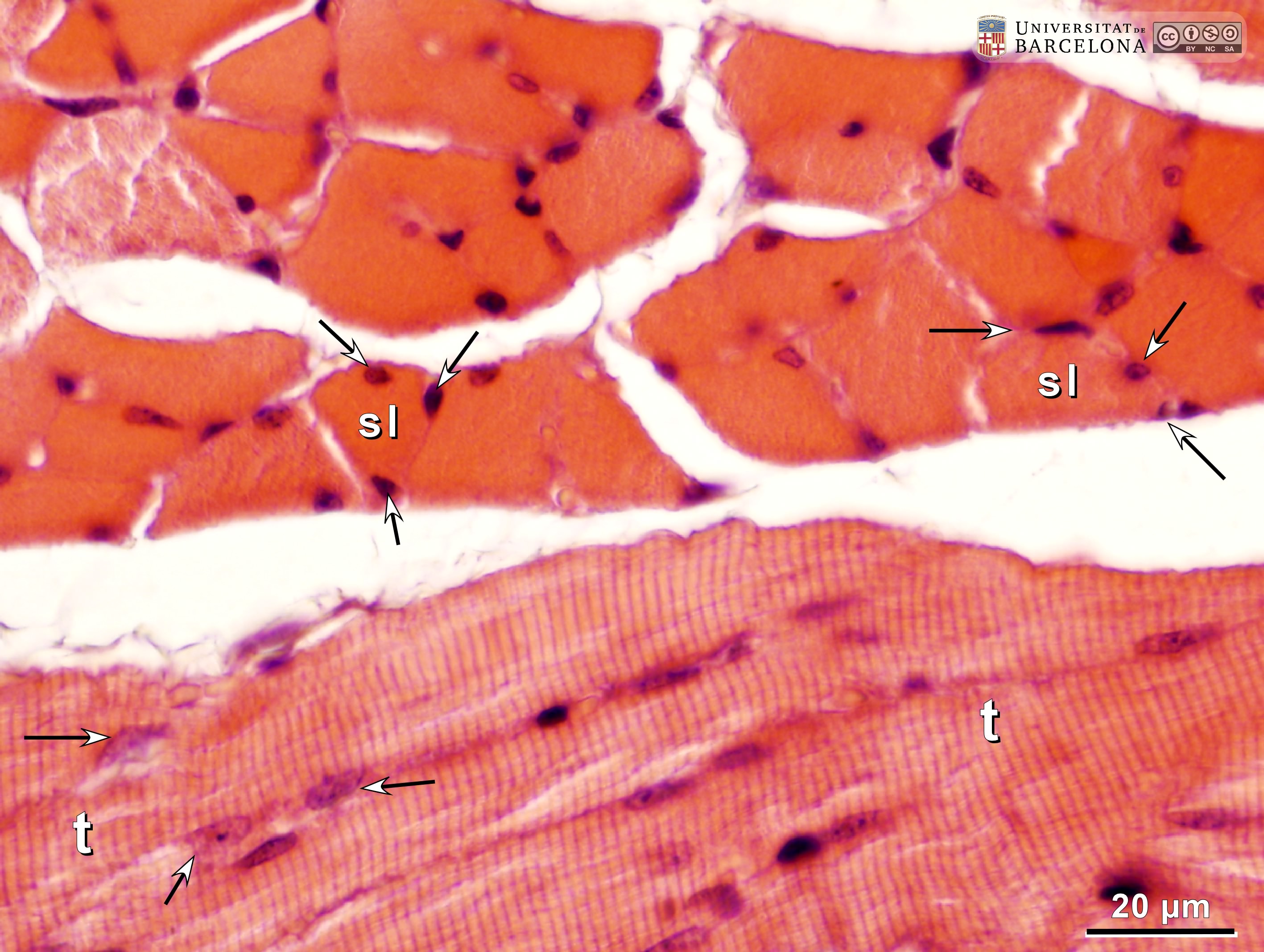 View/Open |
| P_O_tongue_HEosin_50x_38.jpg | Skeletal muscle in the mouse tongue. No labelling. | 754.54 kB | JPEG |  View/Open |
| P_O_tongue_vGieson_2x_b59_b62_collage_fused_LETTERS.jpg | Transverse section of the tip or apex of the mouse tongue. During tissue processing the tip was bent resulting in slight distortion of the midline plane (dashed line). The bulk of the tongue is made of striated muscles stained brown with van Gieson trichrome. The superior longitudinal fibre bundles (sl, top) and inferior longitudinal bundles (il, bottom) are seen in the periphery, while the transverse muscles (t) fill the central part. Connective tissue is present between the fibre bundles, and its collagen fibres are stained dark pink. Several arteries (ar), veins (v) and nerve branches can be seen within this pink background. The lingual mucosa, on the surface, consists of a thick epithelium (ep) with conical protrusions – the papillae (asterisks) – and a deep, thinner layer of connective tissue – the lamina propria (lp) – with pink-stained collagen fibres, more conspicuous in the ventral part of the tongue. The lamina propria is continuous with the connective tissue between the muscle bundles. Paraffin section, van Gieson trichrome. | 808.88 kB | JPEG |  View/Open |
| P_O_tongue_vGieson_2x_b59_b62_collage_fused.jpg | Transverse section of the tip or apex of the mouse tongue. No labelling. | 764.61 kB | JPEG | 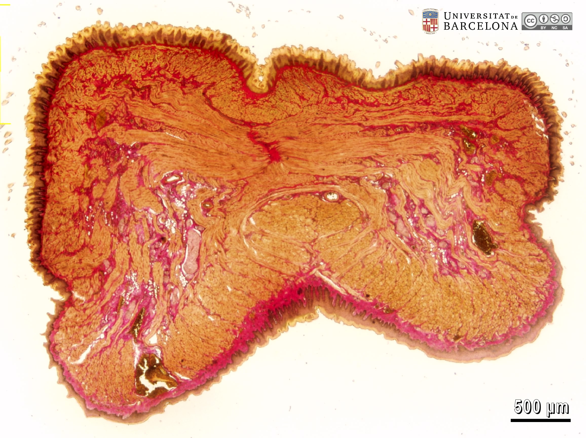 View/Open |
| P_O_tongue_vGieson_4x_b72_b78_collageB_LETTERS.jpg | Transverse section of the mouse tongue close to the apex (collage of micrographs allowing moderate zoom). The midline (slightly distorted) is marked by the dashed line. The epithelium (ep) is stained light brown on the surface (layer of dead cells) and dark brown in deeper layers. On the dorsal and lateral surfaces, conical projections are observed: the filiform papillae (asterisks). The lamina propria (lp) is located beneath the epithelium. This is a layer of connective tissue rich in collagen fibres stained pink with acid fuchsin. Skeletal muscle fibres are stained brown. Some are cut transversally: the superior longitudinal (sl) and inferior longitudinal (il) muscles. The vertical muscle bundles (ve) are thin and cut longitudinally, as are the massive transverse muscles (t), which are anchored to the centre of the tongue (dashed circle). Connective tissue (stained pink) including adipose tissue (a) fills the gaps between the fibre bundles and typically accommodates arteries (ar), veins (v) and nerves (n), which are symmetrically distributed on each side of the midline. Paraffin section, van Gieson trichrome. | 2.46 MB | JPEG | 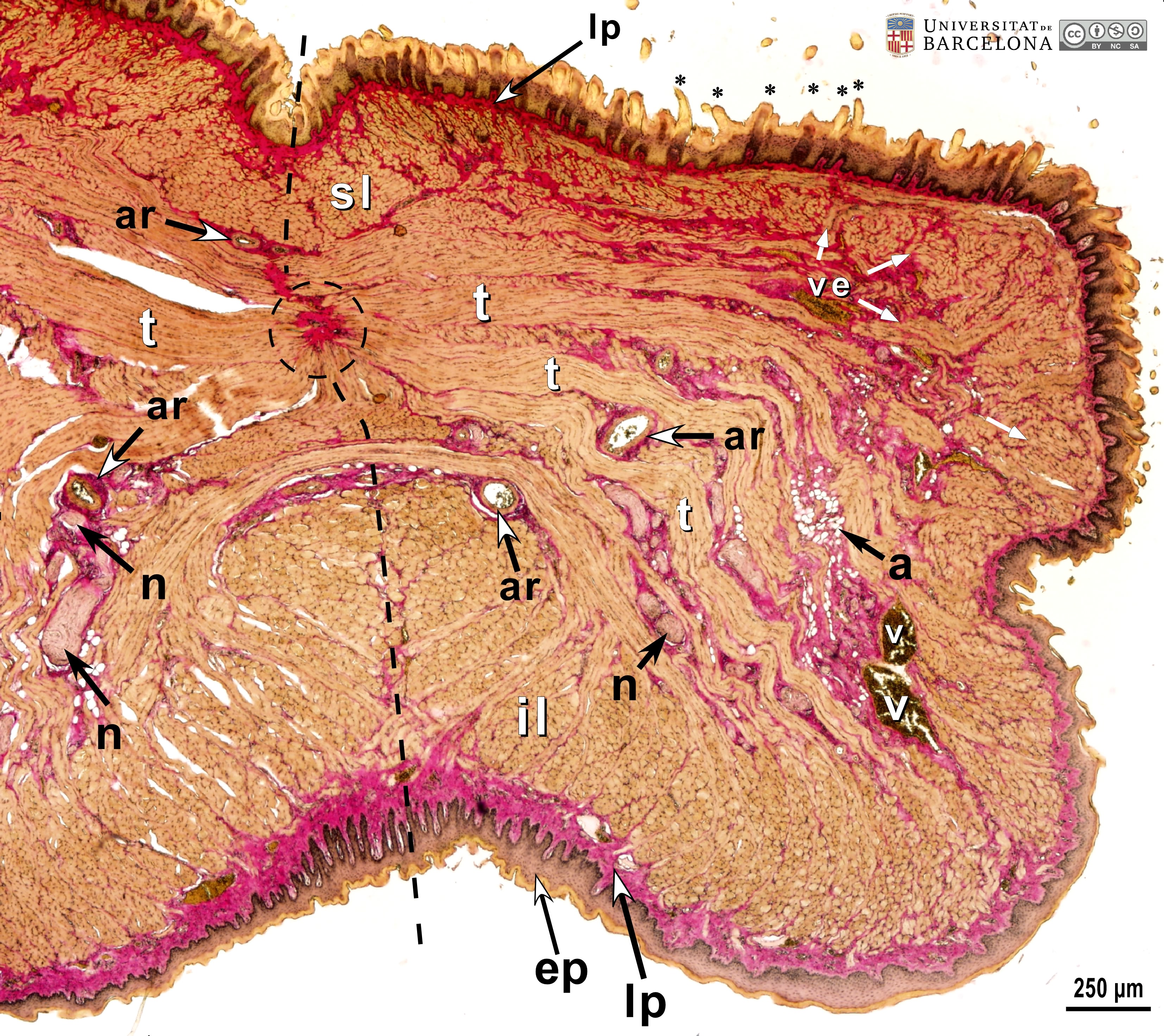 View/Open |
| P_O_tongue_vGieson_4x_b72_b78_collageB.jpg | Transverse section of the mouse tongue close to the apex (collage of micrographs allowing moderate zoom). No labelling. | 2.41 MB | JPEG | 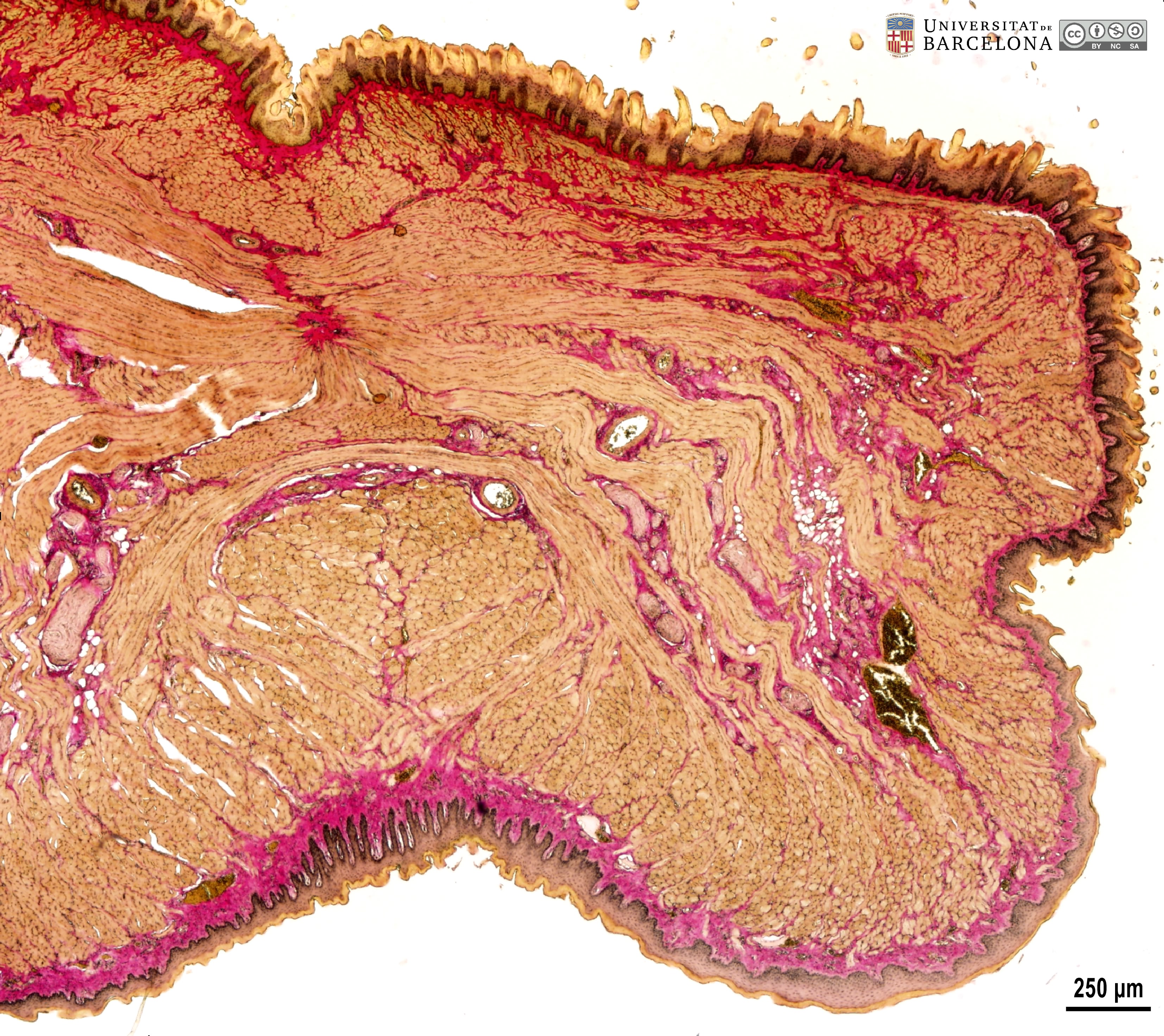 View/Open |
| P_O_tongue_vGieson_4x_b66_b68_collage_trimmed2_LETTERS.jpg | Transverse section of the mouse tongue (collage of micrographs allowing moderate zoom). The epithelium (ep) on the dorsal (top) and lateral surfaces is thick and uneven with conical protrusions or papillae (asterisks). On the ventral surface, the epithelium is more even and thinner. Deeper layers of the epithelium are dark brown, while the superficial layer is lighter (the layer of dead cells). Beneath the epithelium, the collagen fibres in the lamina propria (lp) stain deep pink. In the ventral portion of the tongue, the lamina propria is wider and adjacent to large veins (v). Muscle fibres are stained brown. The superior longitudinal (sl) and inferior longitudinal (il) muscles are distributed in the periphery and are cut transversally here. Vertical muscle (ve) bundles are thin and intercalated with the longitudinal muscles. The core of the tongue is occupied by the thick bundles of the transverse muscles (t). Interspersed with the fibre bundles, there is connective tissue stained pink that surrounds nerves (n), arteries (ar), and veins (v), some of which contain erythrocytes. Paraffin section, van Gieson trichrome. | 1.56 MB | JPEG |  View/Open |
| P_O_tongue_vGieson_4x_b66_b68_collage_trimmed.jpg | Transverse section of the mouse tongue (collage of micrographs allowing moderate zoom). No labelling. | 1.52 MB | JPEG | 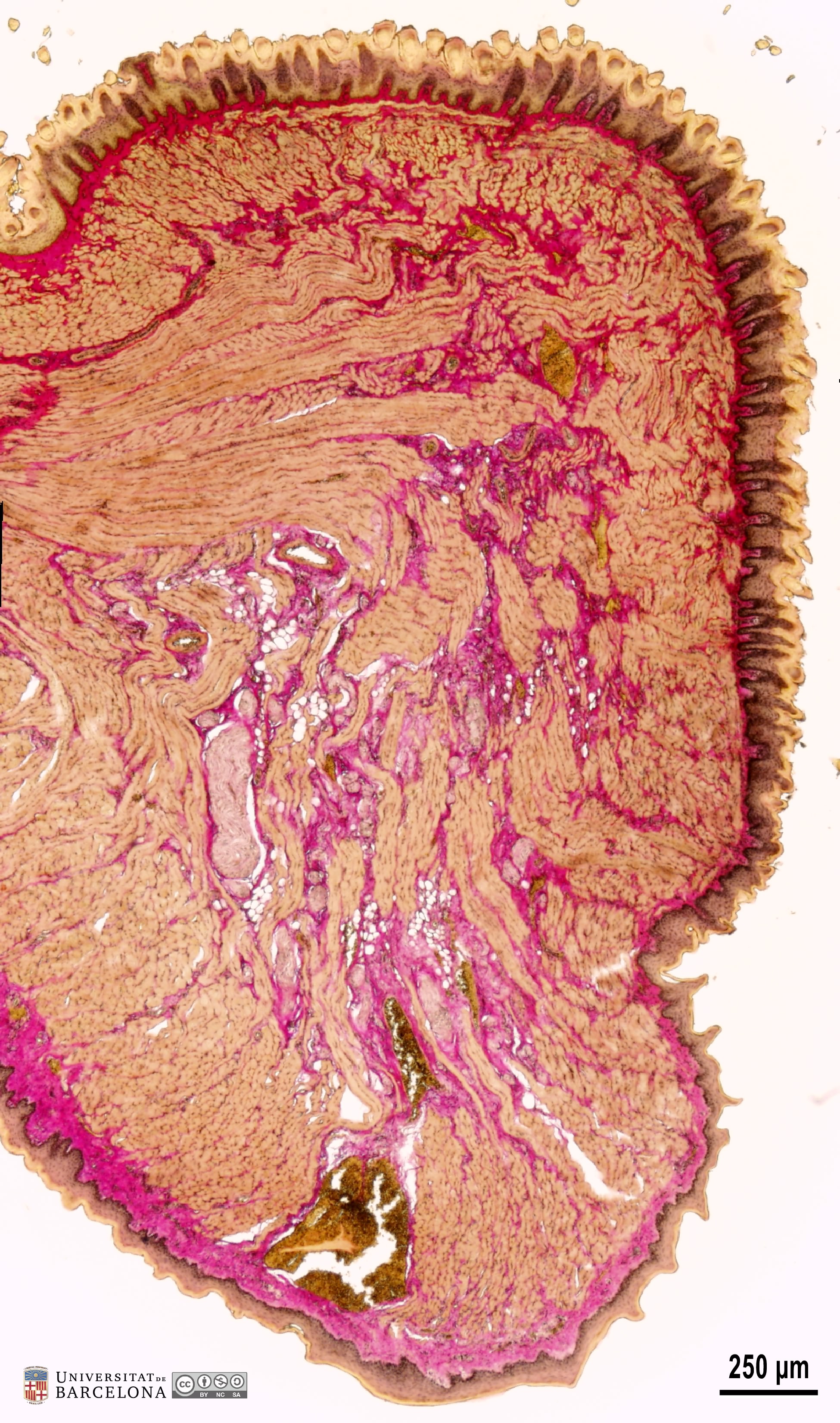 View/Open |
| P_O_tongue_vGieson_4x_b63_LETTERS2.jpg | Detail of the dorsal surface in a mouse tongue. The epithelium (ep) forms conical projections, the filiform papillae (asterisks). The inner layers are stained dark brown, while the superficial layer of dead cells – the stratum corneum – stains pale yellow with picric acid. Beneath the epithelium, there is a layer of connective tissue – the lamina propria (lp) – with collagen fibres stained dark pink or red. The lamina propria forms elevations that push the epithelium upwards (white, empty arrowheads), contributing to the formation of the lingual papillae. The connective tissue also invades deeper layers, enveloping the skeletal (striated) muscle bundles (light brown). Bundles of the superior longitudinal muscles (sl) are cut transversally, while the transverse muscles and the thin bundles of the vertical muscles are cut longitudinally. Deep into the tongue, relatively large arteries (ar), a vein filled with erythrocytes (v), and a nerve (n) can be seen. Paraffin section, van Gieson trichrome. | 1.29 MB | JPEG | 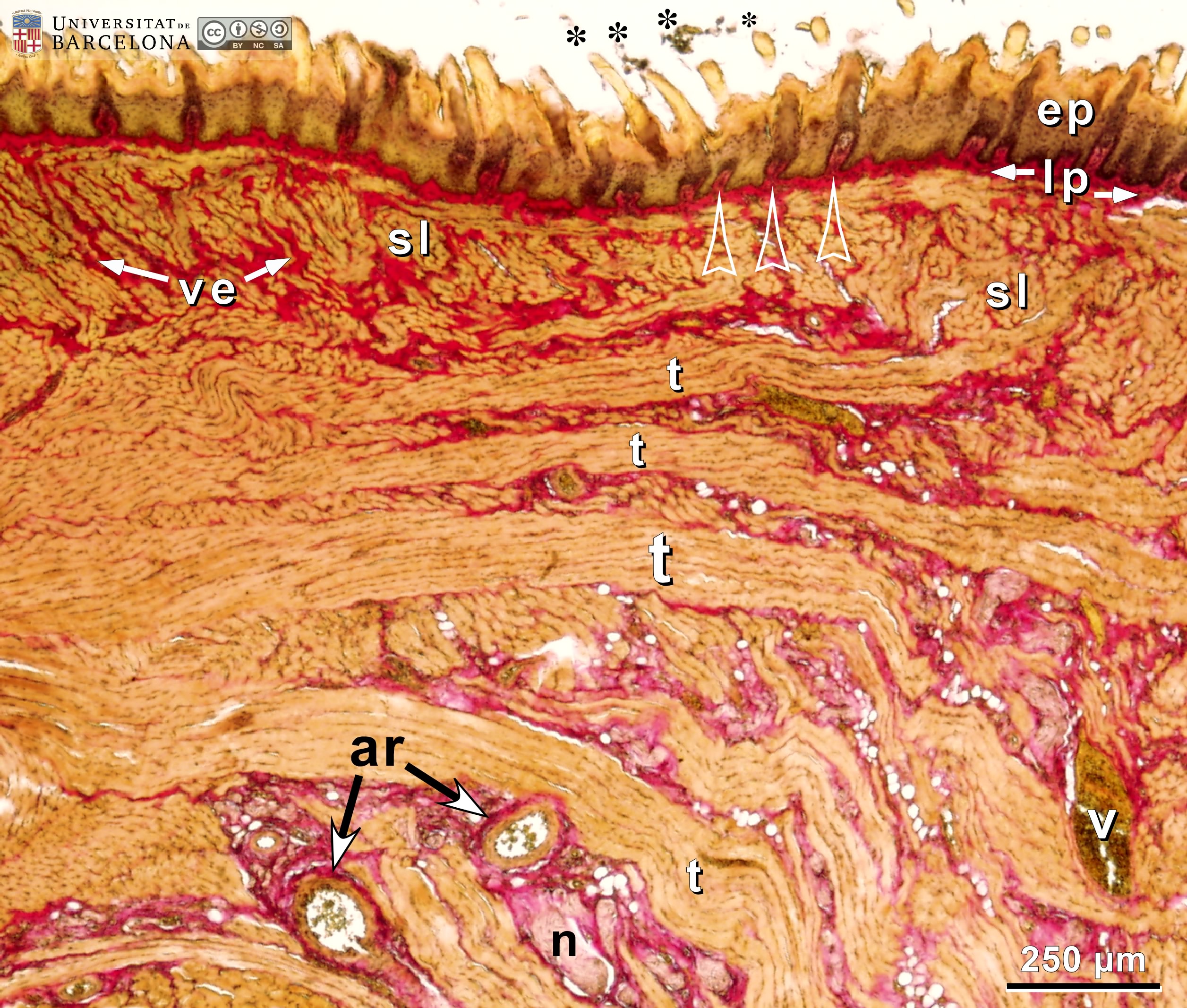 View/Open |
| P_O_tongue_vGieson_4x_b63.jpg | Detail of the dorsal surface in a mouse tongue. No labelling. | 1.26 MB | JPEG | 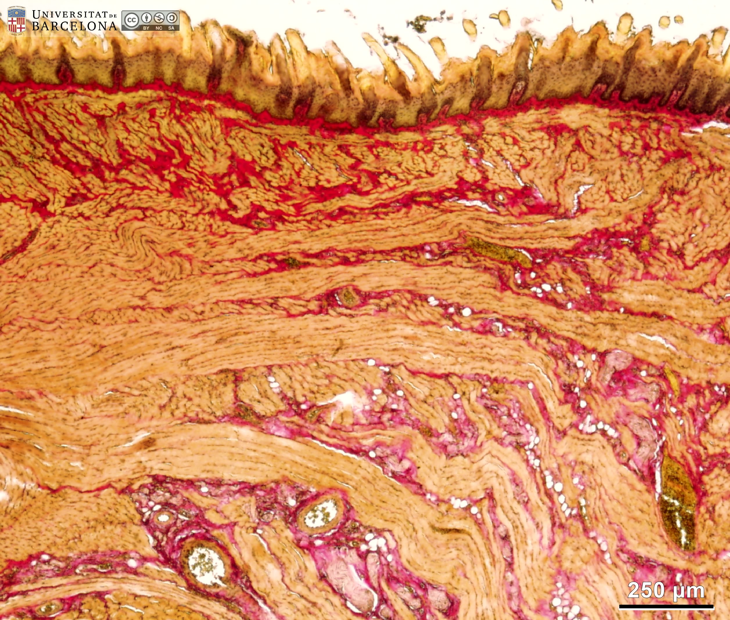 View/Open |
| P_O_tongue_vGieson_4x_b70_b71_collage_LETTERS2.jpg | Ventral surface of a mouse tongue at the midline (dashed segments). Collage of micrographs allowing moderate zoom. The ventral epithelium (ep) has a rather smooth surface, containing an outer layer of dead cells – the stratum corneum – stained light yellow, while deeper strata are brown. Immediately beneath the epithelium, the lamina propria (lp) is thick (thicker than on the dorsal surface), stained pink because of the collagen fibres, and protruding downwards into the epithelium (arrowheads), resembling the dermal papillae found in skin. Deeper into the tongue, bundles of the inferior longitudinal muscles (il) were sectioned transversally, and those of the tranverse muscles longitudinally. Immersed in the connective tissue between the muscular bundles, arteries (ar) and nerves (n) are symmetrically distributed. Paraffin section, van Gieson trichrome. | 1.07 MB | JPEG | 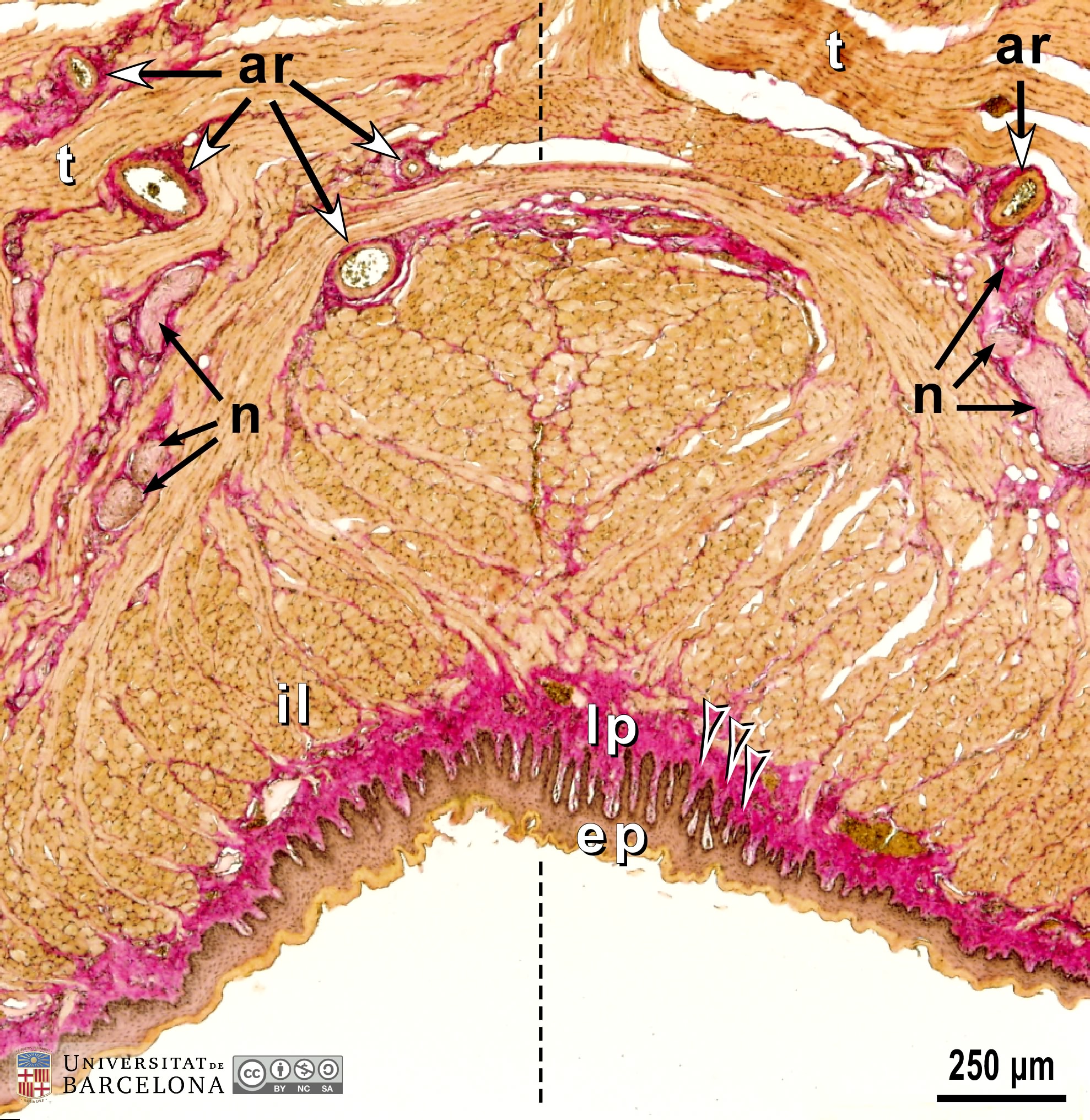 View/Open |
| P_O_tongue_vGieson_4x_b70_b71_collage.jpg | Ventral surface of a mouse tongue at the midline. No labelling. | 1.05 MB | JPEG | 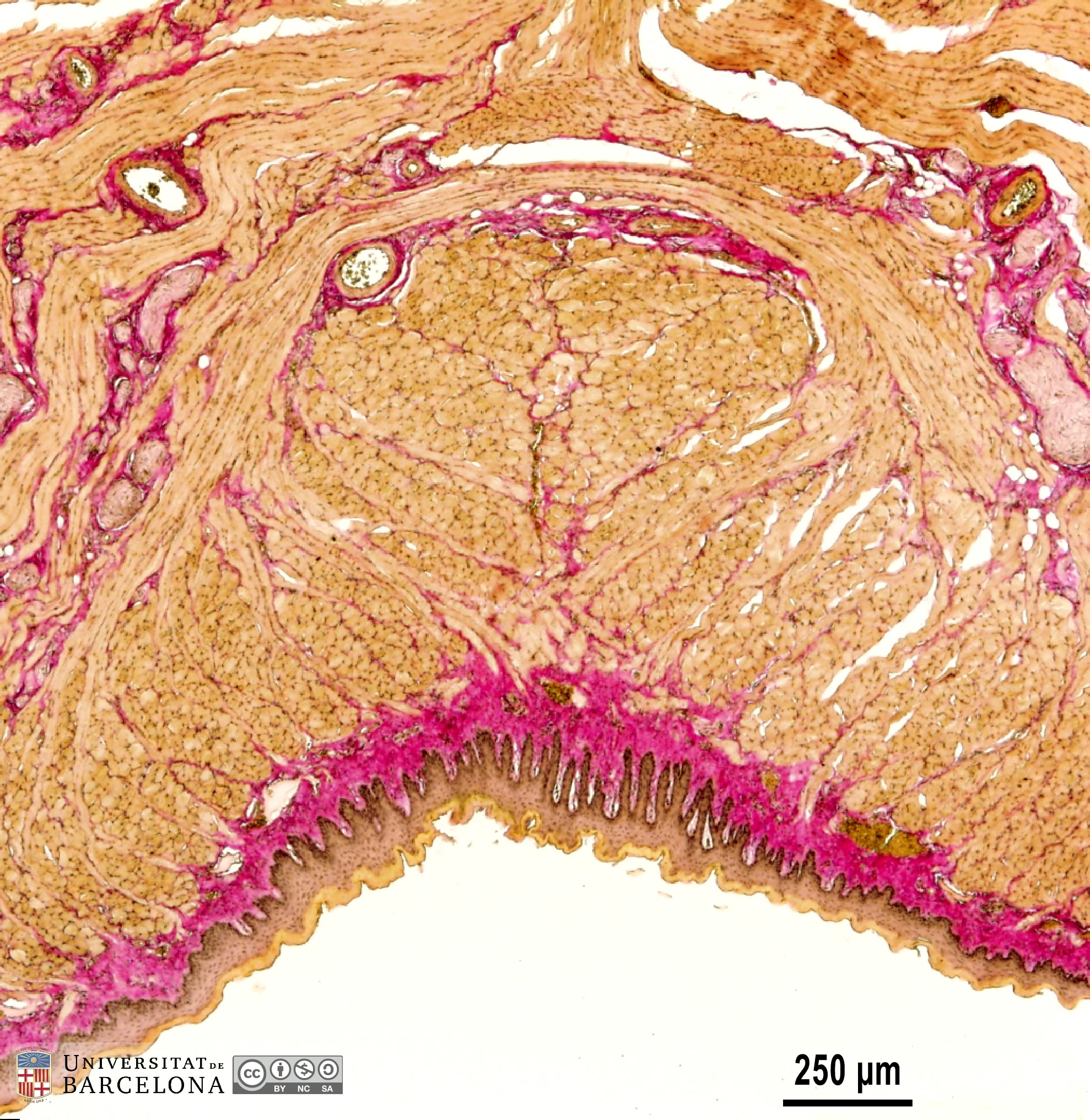 View/Open |
| P_O_tongue_vGieson_20x_64_LETTERS.jpg | Dorsal surface of a mouse tongue. The mucosa consists of an epithelium (ep) stained brown, and a thin lamina propria with collagen fibres stained pink. Deep to the mucosa, bundles of skeletal (striated) muscle are stained light brown. A keratinised stratified squamous epithelium is seen at the surface and contains the typical layers: stratum basale (bas) or germinative layer, with dark brown cell nuclei, stratum spinosum (sp) with moderately stained cell nuclei, although cells lack the spiny appearance, stratum granulosum (gra) with dark brown keratohyaline granules (not neatly visible at this magnification), and stratum corneum (cor), stained light brown or yellow, and composed of anucleated dead cells. The lamina propria (lp) contains dispersed cells stained brown, and collagen fibres in the extracellular matrix, stained dark pink with acid fuchsin. The lamina propria widens at regular intervals (white stars) and pushes the epithelium upwards, forming conical projections: the filiform papillae (asterisks). Paraffin section, van Gieson trichrome. | 794.72 kB | JPEG | 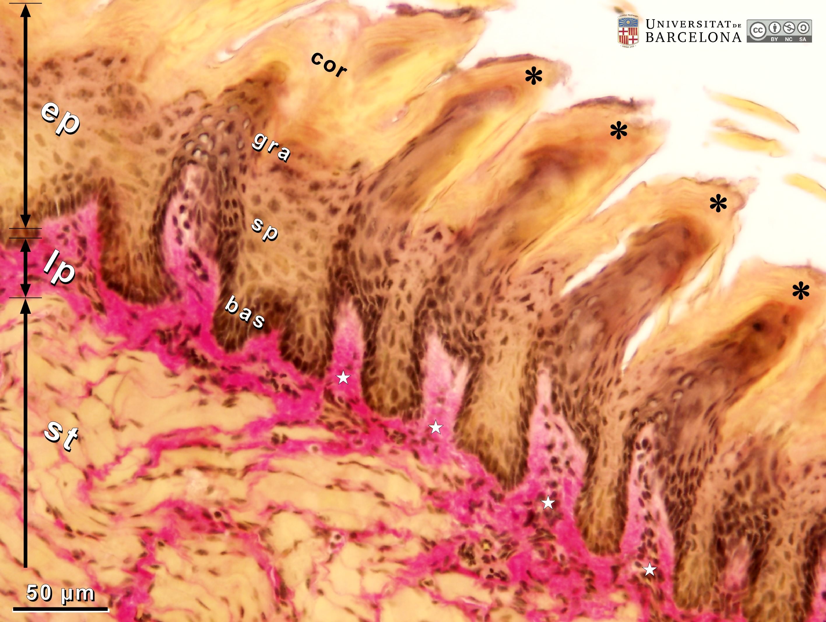 View/Open |
| P_O_tongue_vGieson_20x_64.jpg | Dorsal surface of a mouse tongue. No labelling. | 752.78 kB | JPEG |  View/Open |
| P_O_tongue_vGieson_20x_69_LETTERS.jpg | Ventral surface of a mouse tongue in its anatomical orientation (ventral: at the bottom). Deep to the epithelium (ep) there is a distinct lamina propria (lp) stained pink, and deeper to it the striated muscle bundles (st). The epithelium forms a smooth overlay and includes several layers: stratum corneum (cor) of dead cells accumulating at the surface, stratum granulosum (gra) where dark keratohyaline granules can be seen when enlarging the image, stratum spinosum (sp) with elongated cell nuclei, and an undulating stratum basale (bas) with dark – almost black – cell nuclei. The lamina propria contains heavily stained collagen fibres, scattered cells, and numerous blood vessels containing erythrocytes with a yellowish tint, due to picric acid staining. In the muscular zone, the bundles of the inferior longitudinal muscle (il) are wrapped in thin layers of connective tissue containing collagen fibres stained pink (empty arrowheads). Paraffin section, van Gieson trichrome. | 842.37 kB | JPEG | 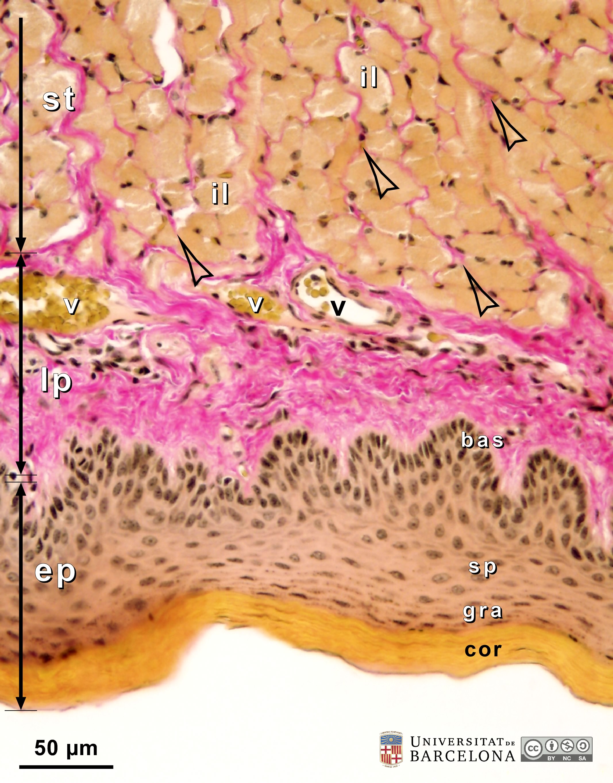 View/Open |
| P_O_tongue_vGieson_20x_69.jpg | Ventral surface of a mouse tongue in its anatomical orientation (ventral: at the bottom). No labelling. | 799.47 kB | JPEG | 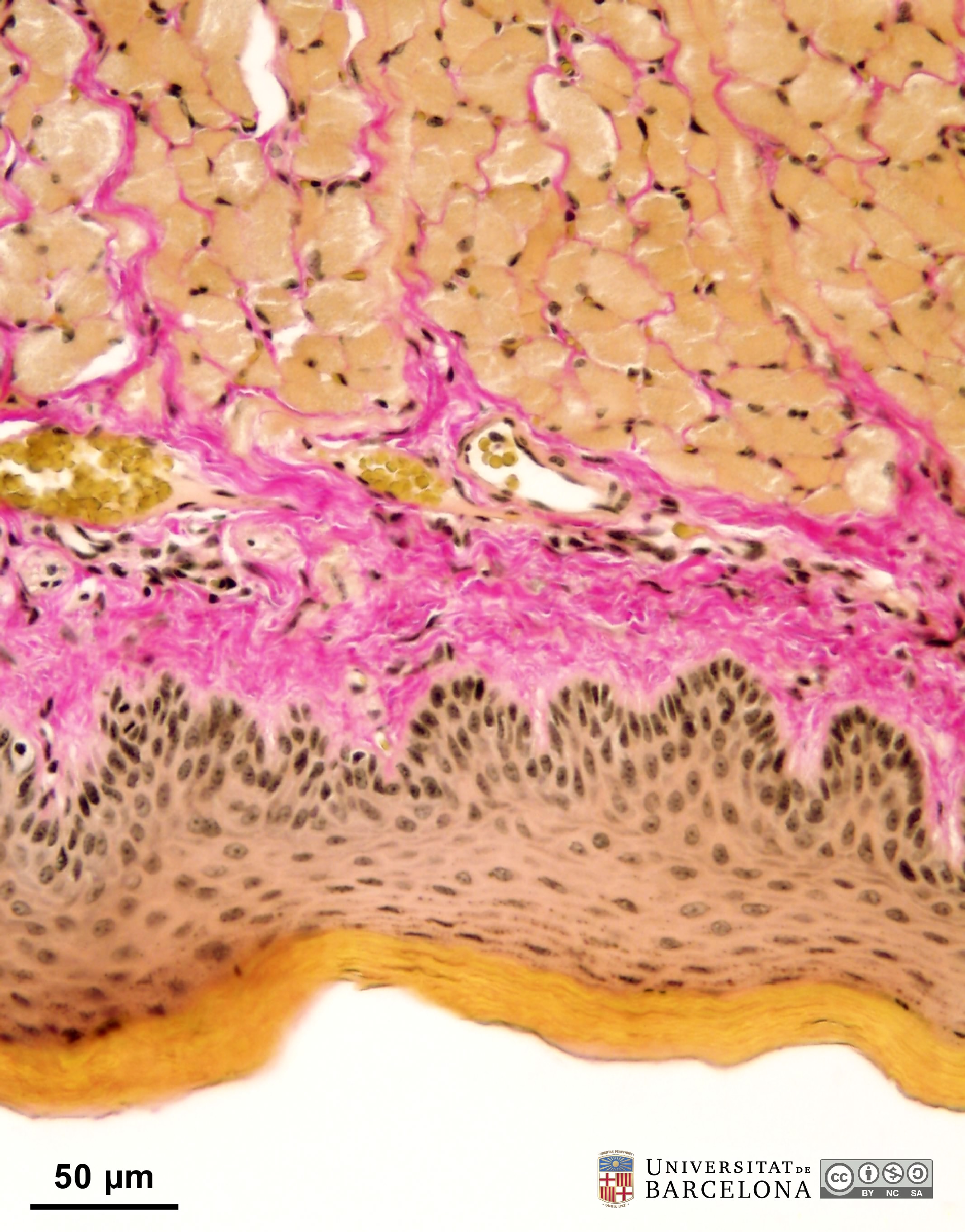 View/Open |
| P_O_tongue_vGieson_50x_85_LETTERS.jpg | Filiform papilla on the dorsal surface of the mouse tongue. A thin lamina propria (lp) widens and forms an elevation. The stratum basale (bas) in the epithelium is pushed upwards, and the whole epithelium, including the lightly stained stratum corneum (cor), forms a conical projection (asterisk). Paraffin section, van Gieson trichrome. Empty arrowheads, collagen fibres; gra, epithelial stratum granulosum; sp, epithelial stratum spinosum; st, striated (skeletal) muscle. | 673.87 kB | JPEG | 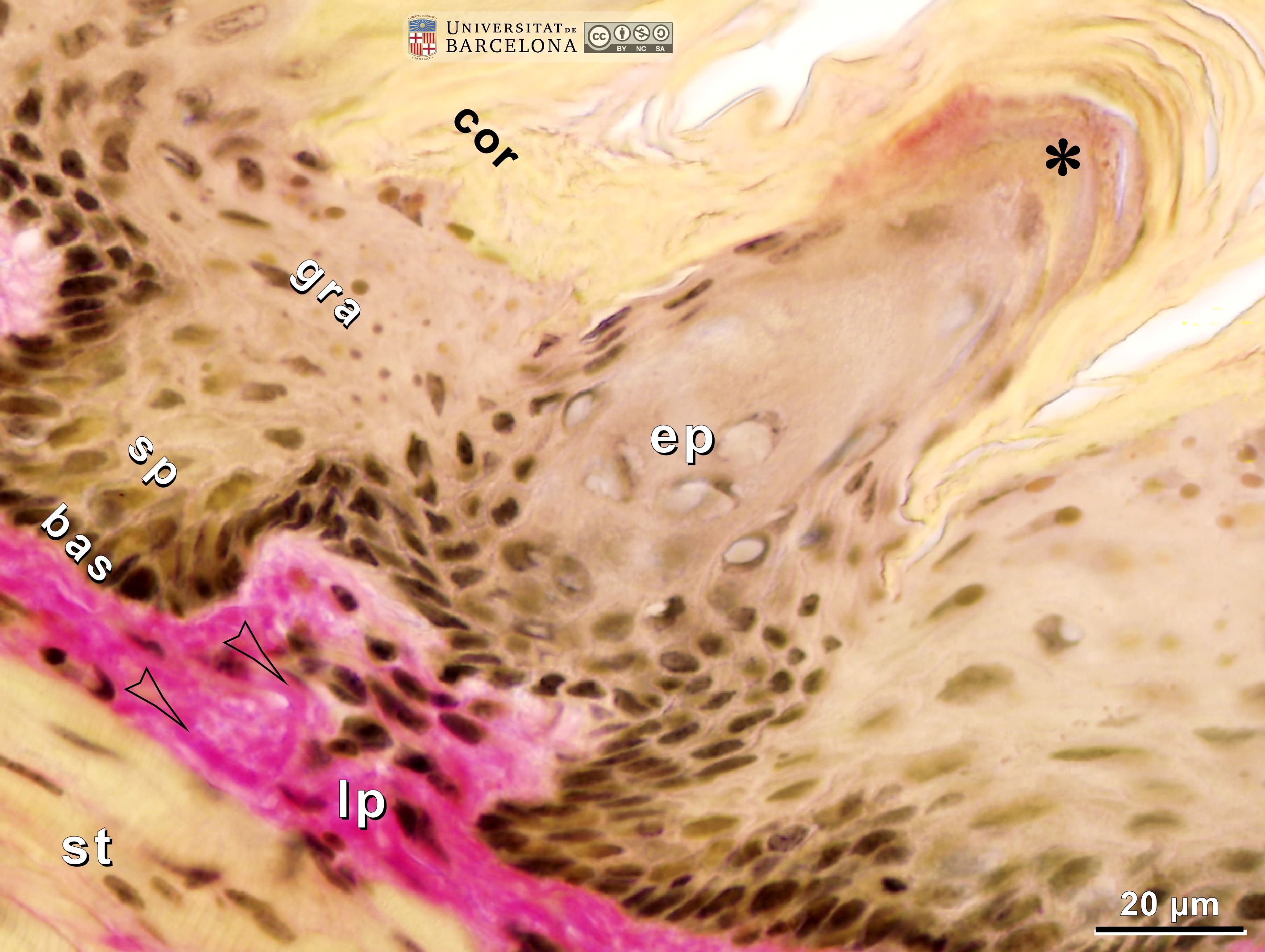 View/Open |
| P_O_tongue_vGieson_50x_85.jpg | Filiform papilla on the dorsal surface of the mouse tongue. No labelling. | 638.98 kB | JPEG | 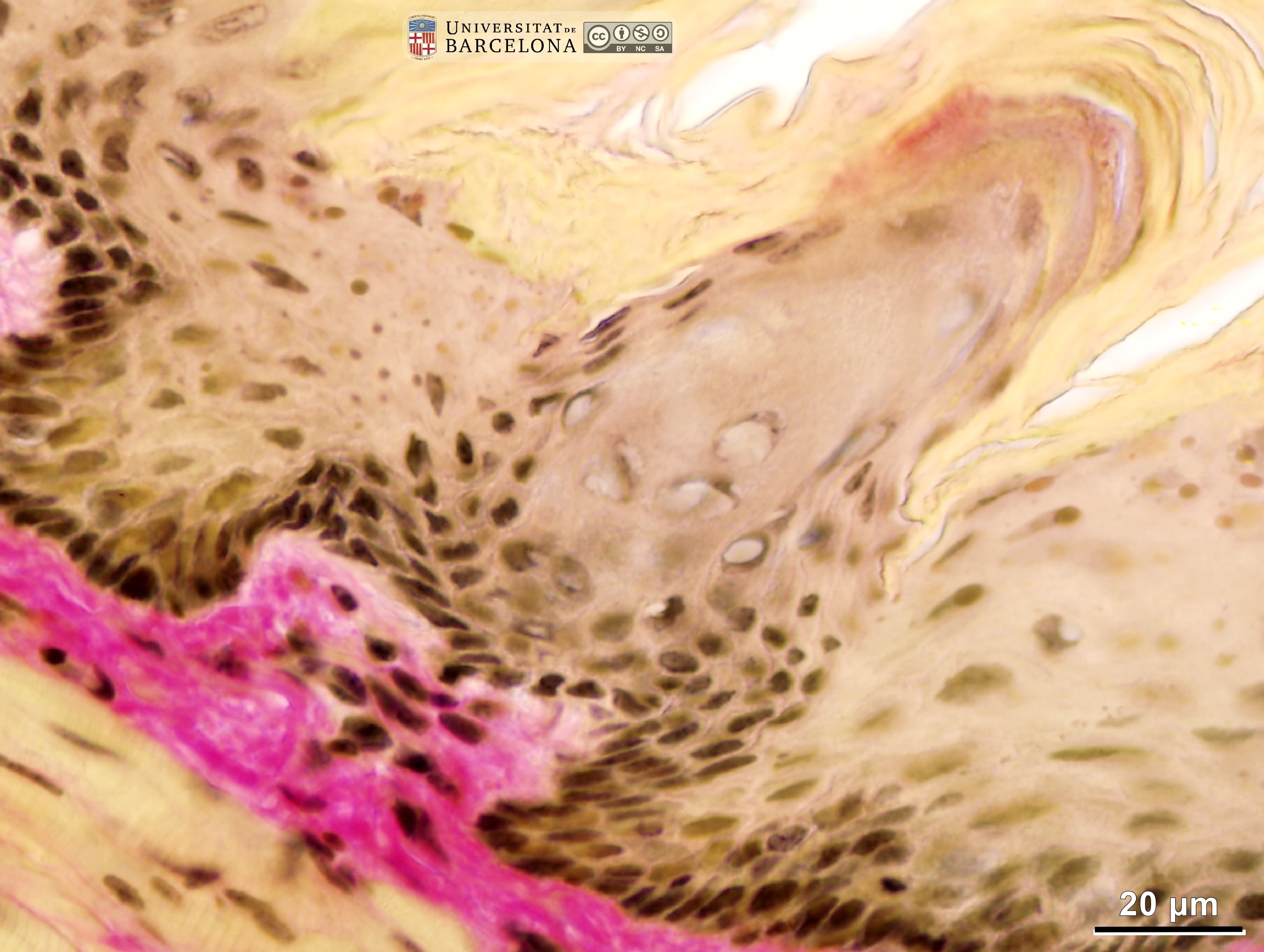 View/Open |
| P_O_tongue_vGieson_50x_81_83_collage_LETTERS.jpg | Ventral surface of the mouse tongue (collage of micrographs allowing detailed zoom; micrograph flipped vertically for layer comparison). The lingual mucosa consists of a stratified epithelium (ep) overlying a connective tissue layer, the lamina propria. Beneath the mucosa, skeletal (striated) muscle fibres (st) can be seen. The epithelium contains the typical layers or strata. Stratum basale (bas) or germinative layer – in contact with the lamina propria –, is thin and contains dark cell nuclei with dark chromatin clumps (observed when zooming in). Stratum spinosum (sp) has cells with oval nuclei which are lightly stained except for a distinct nucleolus. The cells, however, do not look spinous. Stratum granulosum (gra) is easily identified by the numerous small, dark granules in this layer. In this and in deeper layers, cytoplasms are stained light brown or beige, but this changes abruptly at the stratum corneum (cor), which is stained a yellowish tone with picric acid. In this stratum corneum, no cell nucleus is visible, the dead cells are staggered, and their borders are faintly delineated in brown (noticeable when zooming in). The lamina propria contains scattered cells stained dark brown and thick collagen fibres stained deep pink. The bundles of the inferior longitudinal muscles (il) are cut transversally and the striae are not visible: the cells appear pale brown, with dark nuclei (arrows) in the periphery. Blood vessels (v) and pink collagen fibres (empty arrowheads) can be seen between the muscle fibre bundles. Paraffin section, van Gieson trichrome. | 1.15 MB | JPEG | 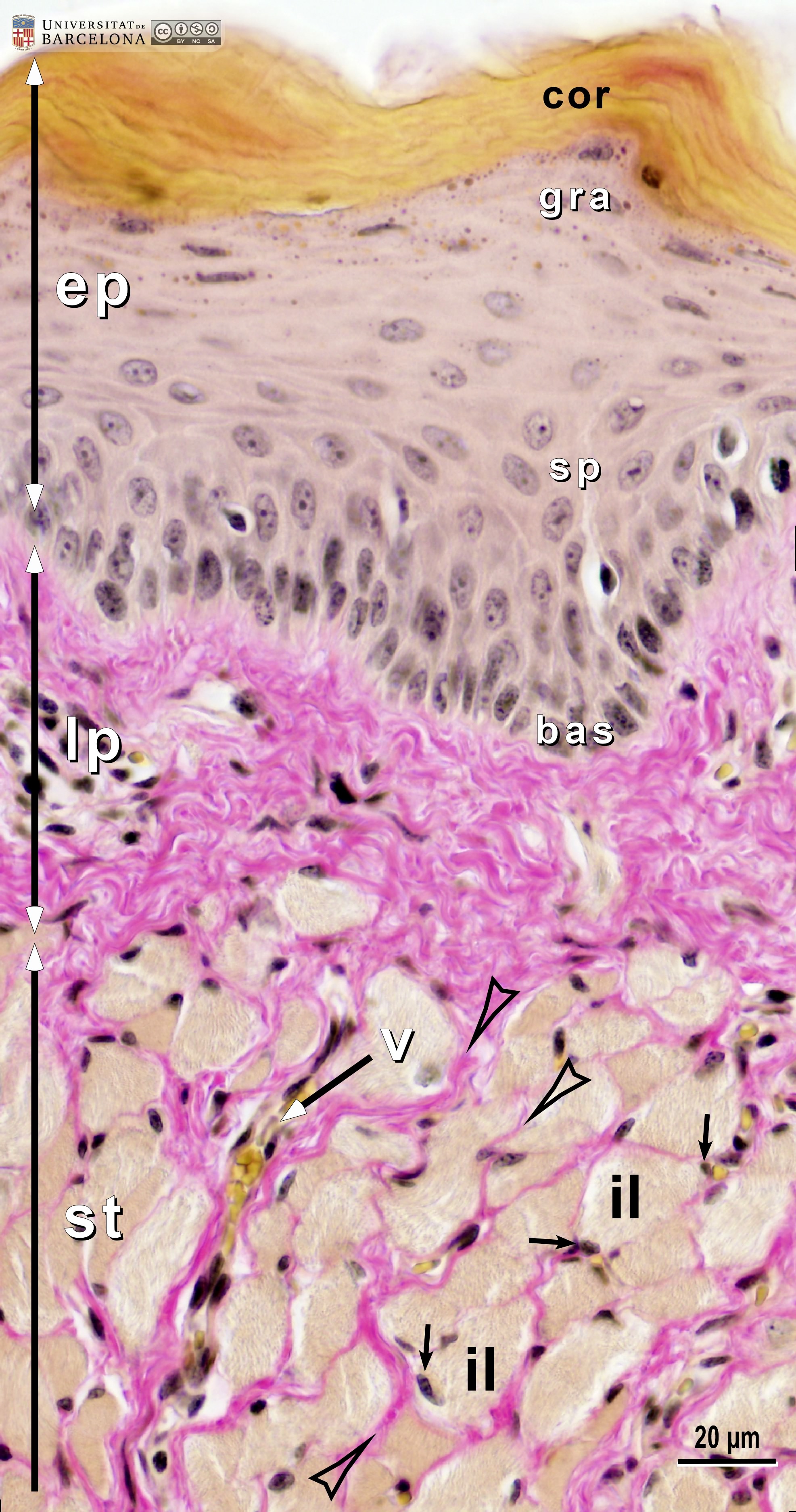 View/Open |
| P_O_tongue_vGieson_50x_81_83_collage.jpg | Ventral surface of the mouse tongue (collage of micrographs allowing detailed zoom; micrograph flipped vertically for layer comparison). No labelling. | 1.1 MB | JPEG | 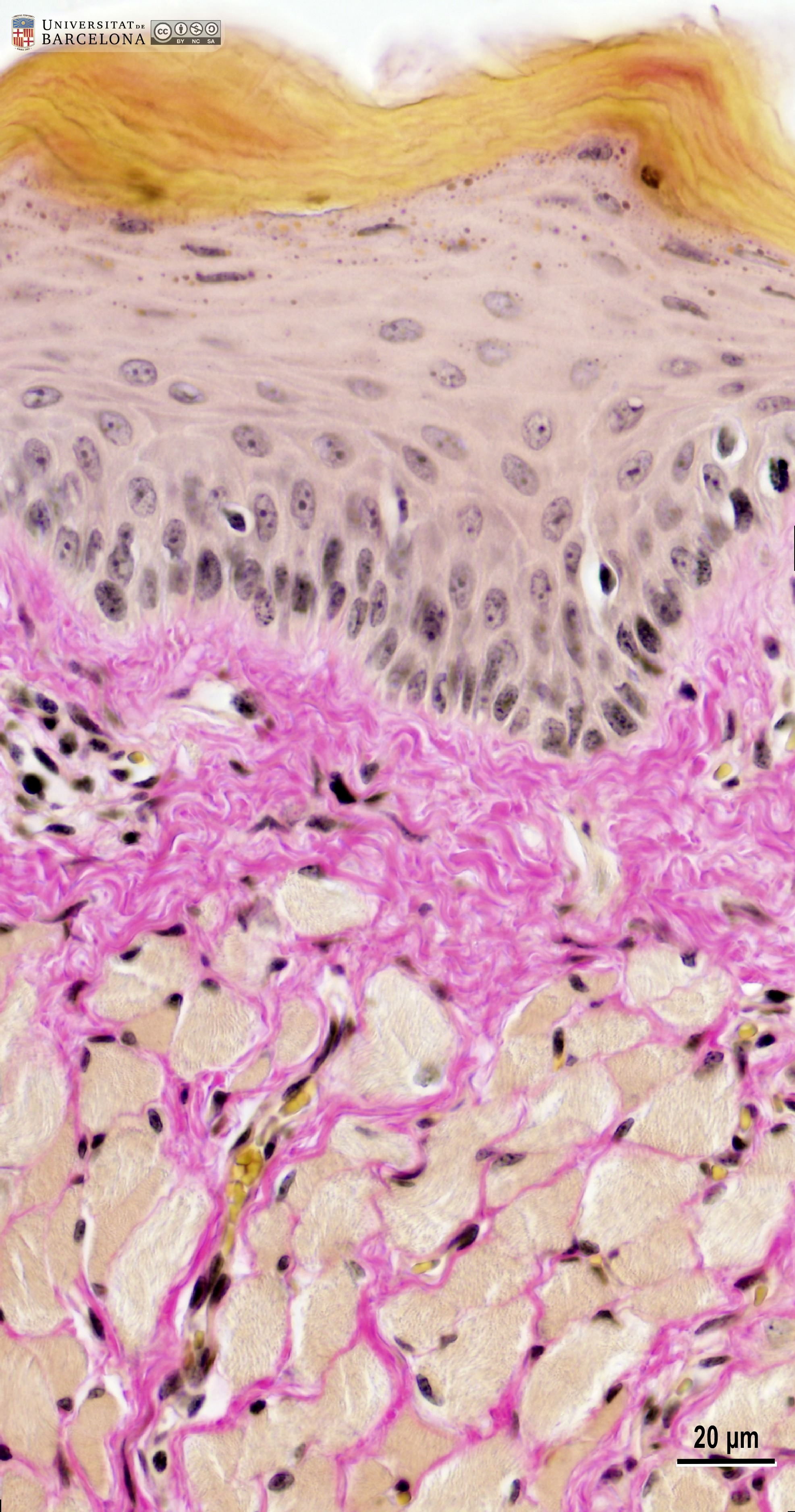 View/Open |
| P_O_tongue_vGieson_20x_91_LETTERS.jpg | Connective tissue deep within the mouse tongue. It fills the space between the skeletal (striated) muscle bundles (st). Thick collagen fibres are stained pink with acid fuchsin, and white adipocytes (a) are unstained and surrounded by thin collagen fibres. Nerve branches (n) and an artery filled with erythrocytes (er) can be seen within the connective tissue. The artery wall shows distinct layers: dark cell nuclei in the tunica intima (ti), smooth muscle fibres stained brown in the tunica media (tm), and a ring of collagen fibres in the tunica adventitia (ta). Paraffin section, van Gieson trichrome. | 908.73 kB | JPEG | 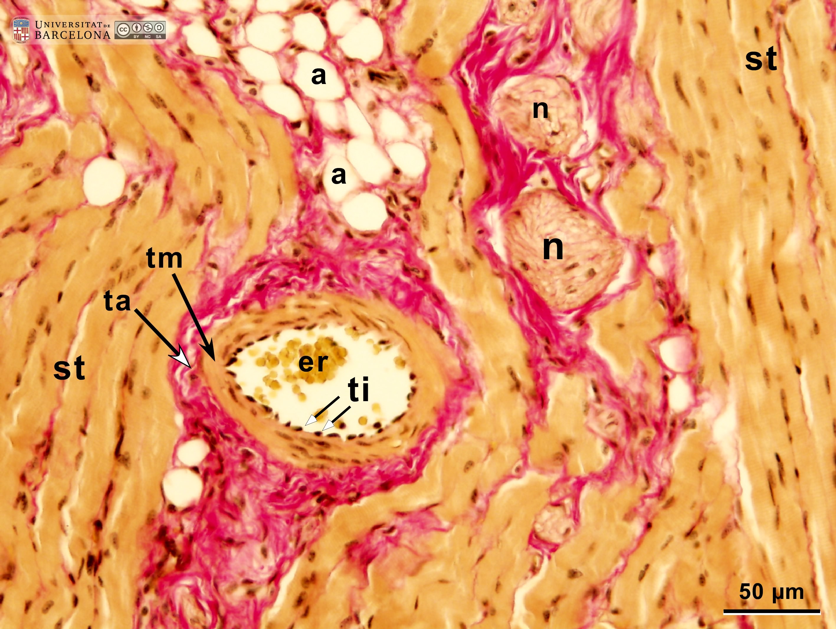 View/Open |
| P_O_tongue_vGieson_20x_91.jpg | Connective tissue deep within the mouse tongue. No labelling. | 884.42 kB | JPEG |  View/Open |
| P_O_tongue_vGieson_50x_76_LETTERS.jpg | Nerves in the mouse tongue. Six nerve branches (1 to 6) are surrounded by thick, pink-stained collagen fibres. Around nerve 2, a thin perineurium (pe) is observed. Within the nerves, cell nuclei (N) are stained dark brown. Small elongated nuclei probably correspond to fibroblasts, and the large round nuclei to Schwann cells. In longitudinal sections (nerves 3 and 5), thin collagen fibres (empty arrowheads) separate pale vertical bands corresponding to myelin sheaths. Paraffin section, van Gieson trichrome. c, capillaries containing erythrocytes. | 911.45 kB | JPEG | 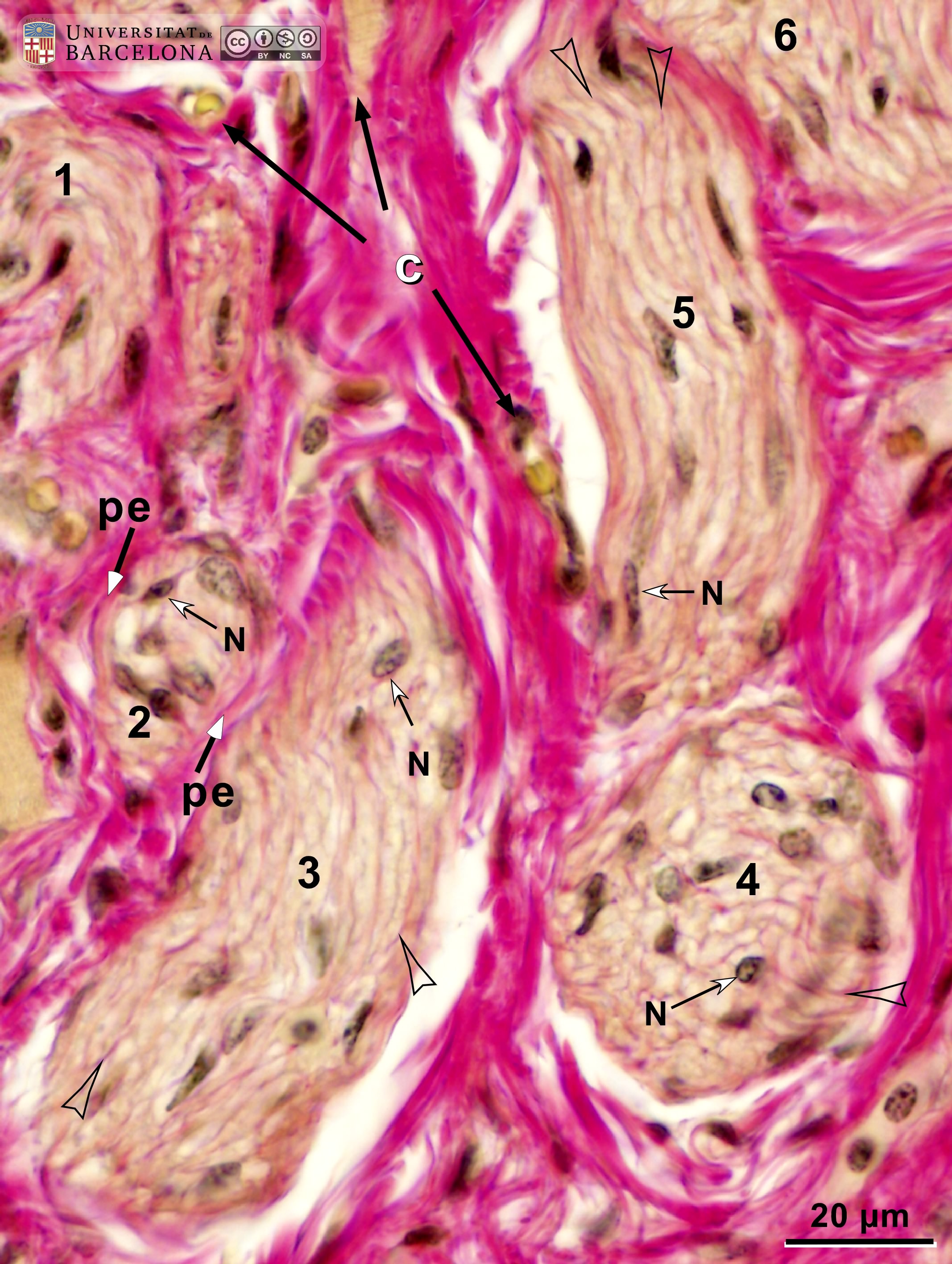 View/Open |
| P_O_tongue_vGieson_50x_76.jpg | Nerves in the mouse tongue. No labelling. | 874.55 kB | JPEG | 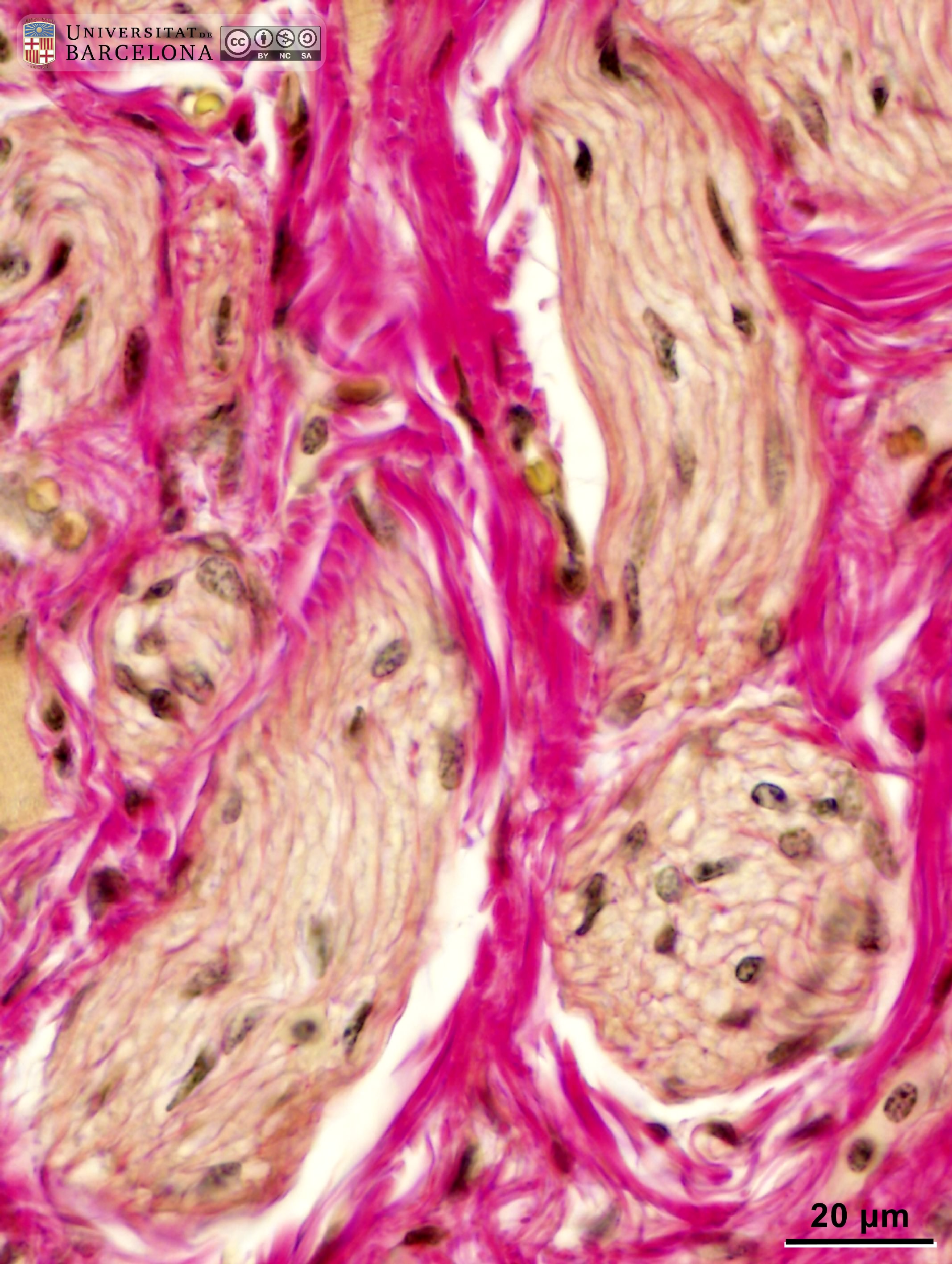 View/Open |
| P_O_tongue_vGieson_50x_77_LETTERS.jpg | Longitudinal section of a nerve in the mouse tongue. The nerve is near skeletal muscle fibres, which are striated (st). The connective tissue contains thick collagen fibres stained pink. The nerve contains thin collagen fibres – the endoneurium (empty arrowheads) – which separate the undulating pale tracks of the myelin sheaths. The whole nerve is enclosed by a thin perineurium (black and white arrows). Paraffin section, van Gieson trichrome. N, cell nuclei. | 903.54 kB | JPEG | 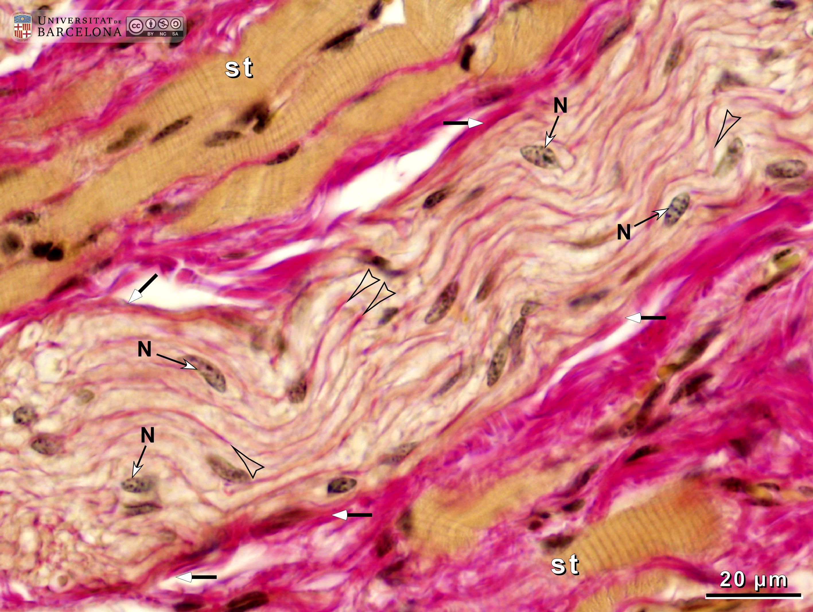 View/Open |
| P_O_tongue_vGieson_50x_77.jpg | Longitudinal section of a nerve in the mouse tongue. No labelling. | 877.1 kB | JPEG | 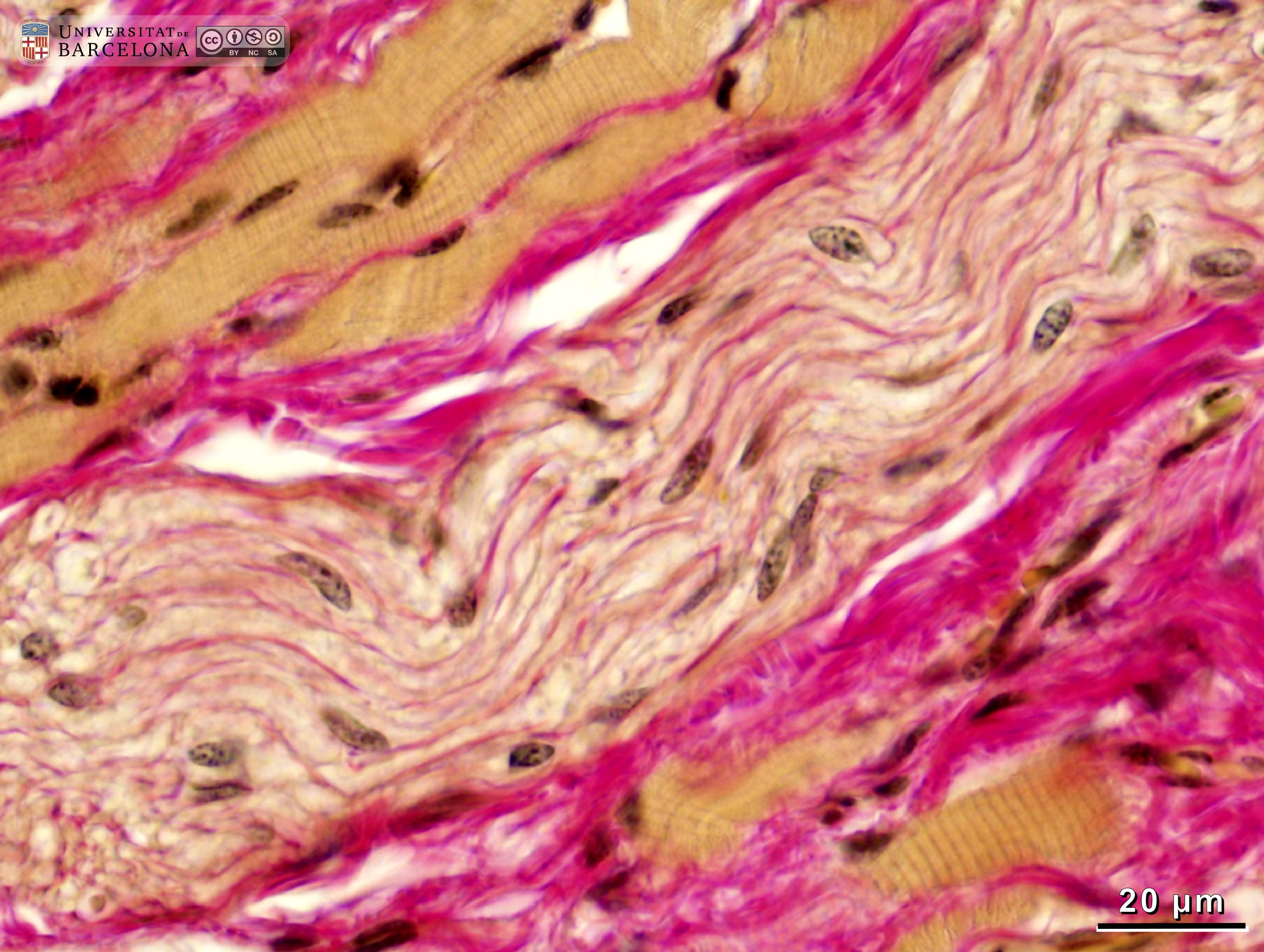 View/Open |
| P_O_tongue_vGieson_50x_101_LETTERS.jpg | Skeletal muscle in the rat tongue. In longitudinal sections (l), the muscle fibres appear striated, with alternating light and dark transverse bands or striae. These striae virtually fill the whole fibre, displacing the nuclei (N) to the periphery. Dark, flattened nuclei (s) probably belong to the satellite cells. The individual muscle fibres are surrounded by thin collagen fibres stained pink: the endomysium (empty arrowheads). Muscle bundles are separated by thicker laminae of collagen fibres, the perimysium (asterisks). Transversally cut muscle fibres (t) have a finely granular content, but myofibrils cannot be observed in this thick paraffin section. As in longitudinal profiles, nuclei are located at the periphery of the fibre and a pink-stained endomysium can also be observed in transverse profiles. van Gieson trichrome. c, capillary. | 882.83 kB | JPEG | 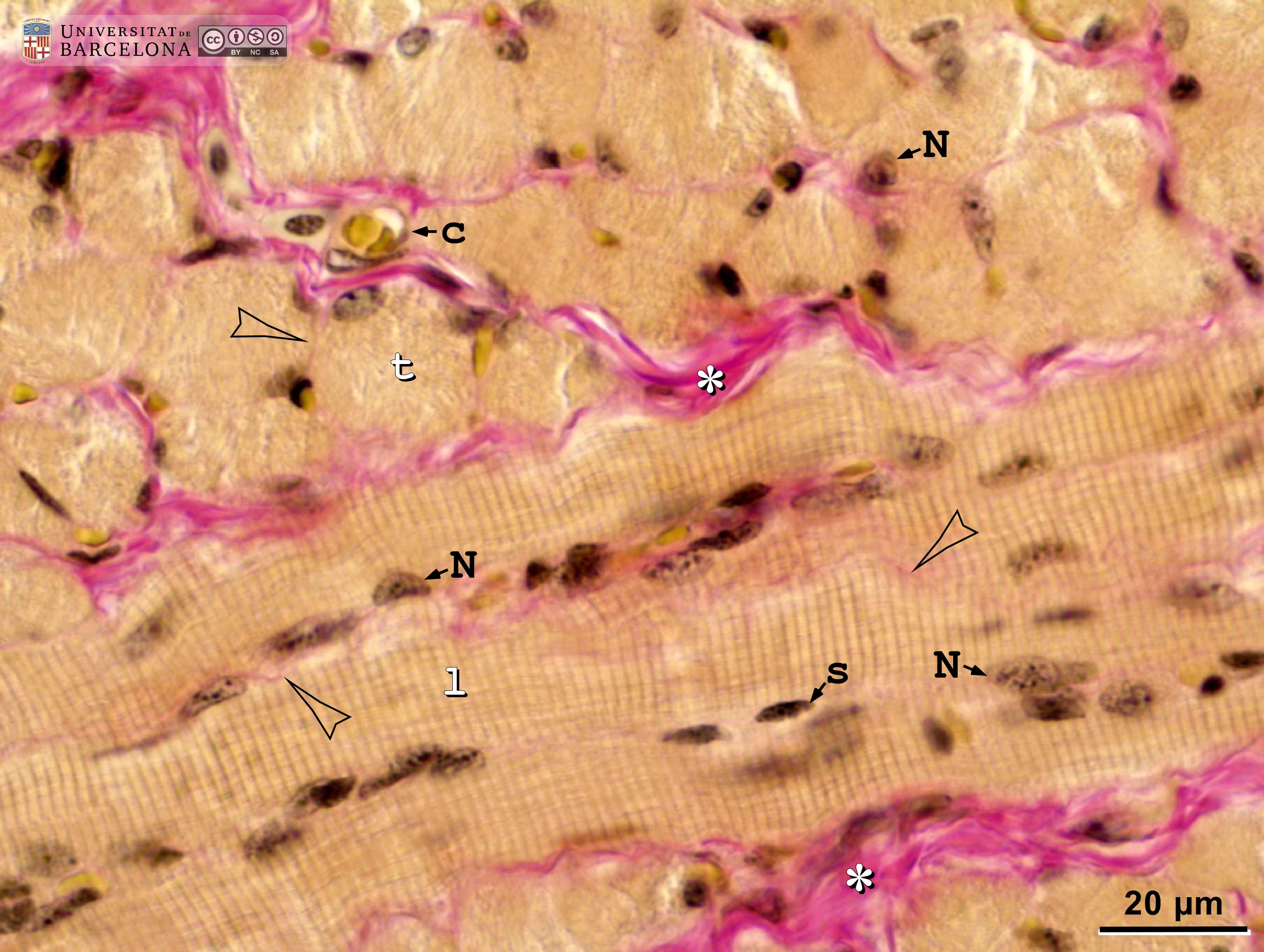 View/Open |
| P_O_tongue_vGieson_50x_101.jpg | Skeletal muscle in the rat tongue. No labelling. | 860.72 kB | JPEG | 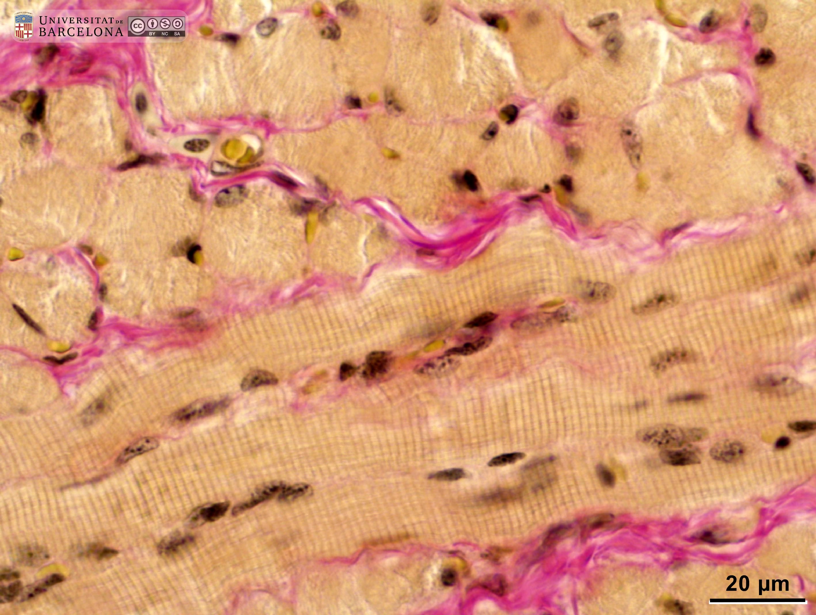 View/Open |
| P_O_tongue_vGieson_50x_112_LETTERS.jpg | Skeletal muscle in the rat tongue. Three muscle fibres are cut longitudinally and cross the picture from left to right (and a fourth fibre at the upper right border). These muscle fibres contain light and dark transverse bands (striae) reflecting the regular organisation of myofibrils. Cell nuclei (N) are located at the periphery of the fibre. Each muscle fibre is surrounded by thin collagen fibres stained pink – the endomysium – (empty arrowheads), and muscle bundles are covered with thicker layers of connective tissue – the perimysium – (asterisks). Transversally cut fibres (t) show a granular cytoplasm lacking striae, while nuclei are located at the periphery of the fibre, as in longitudinal sections. The thin endomysium is also visible around transversally cut fibres (arrowheads). Paraffin section, van Gieson trichrome. | 1.05 MB | JPEG | 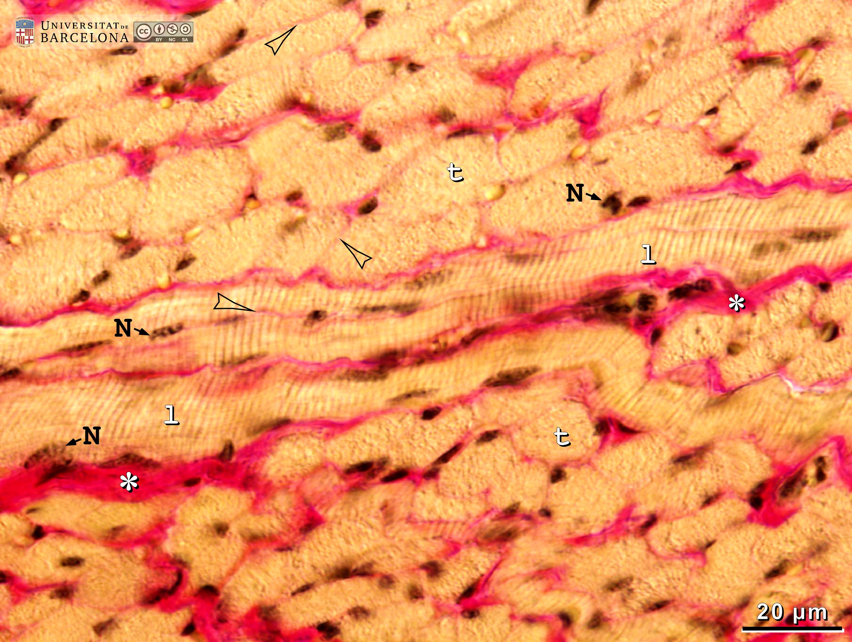 View/Open |
| P_O_tongue_vGieson_50x_112.jpg | Skeletal muscle in the rat tongue. No labelling. | 1.04 MB | JPEG | 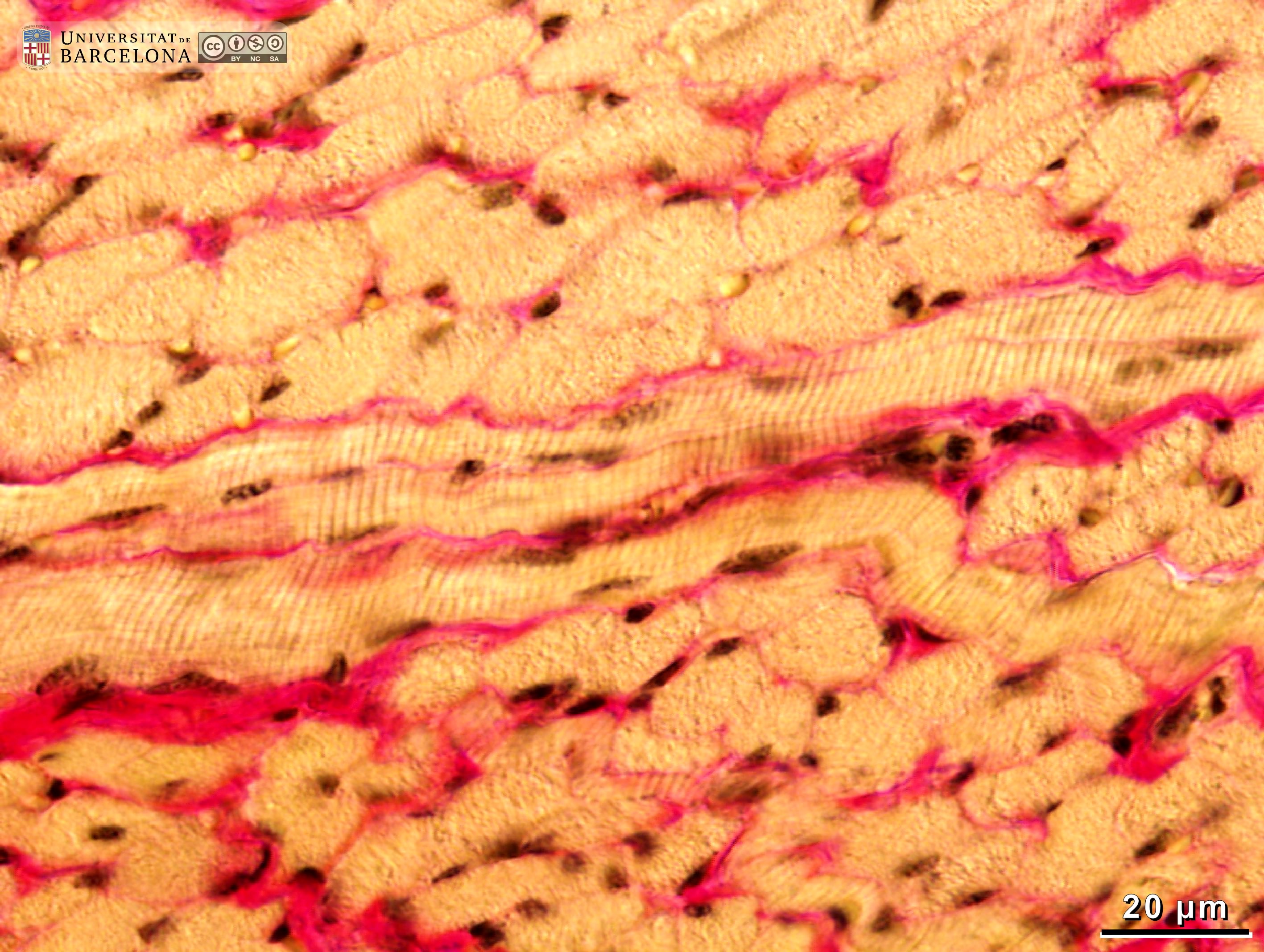 View/Open |
| P_O_tongue_vGieson_50x_104_LETTERS.jpg | Skeletal muscle in the rat tongue. Two muscular bundles are cut longitudinally (l) and run diagonally at the top and bottom of the micrograph. Transversally cut fibres (t) fill the space between them. Longitudinal fibres contain light and dark transverse bands or striae that appear to fill the whole cytoplasm, and nuclei (N) are pushed to the periphery. In transverse fibres, no stria can be observed but nuclei are similarly displaced to the periphery. Both types of fibres are surrounded by delicate collagen fibres stained pink (empty arrowheads). Paraffin section, van Gieson trichrome. | 1.05 MB | JPEG | 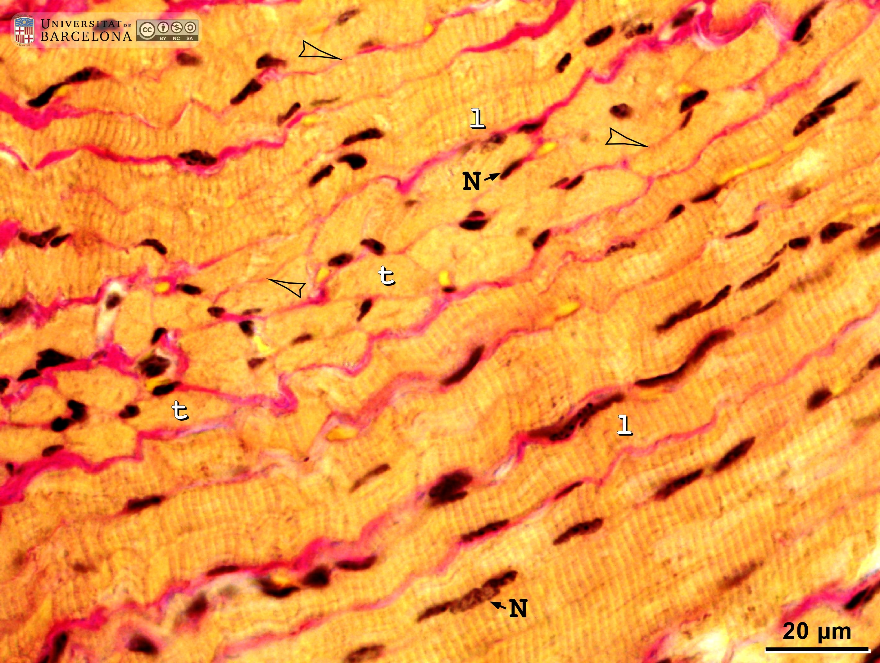 View/Open |
| P_O_tongue_vGieson_50x_104.jpg | Skeletal muscle in the rat tongue. No labelling. | 1.04 MB | JPEG | 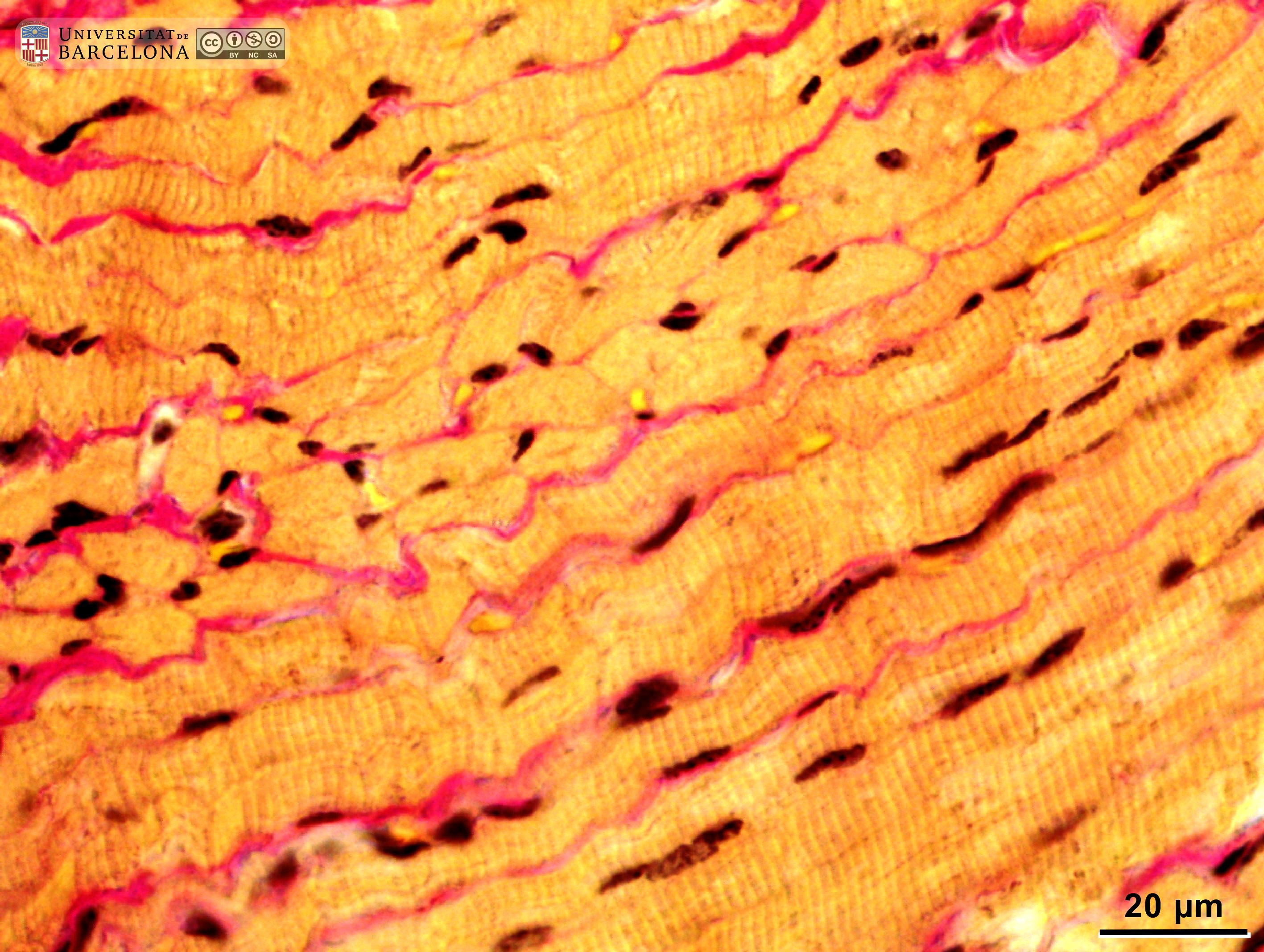 View/Open |
| P_O_tongue_semithin_2um_4x_b087_LETTERS.jpg | Dorsal surface of the rat tongue. The epithelium forms conical projections: the filiform papillae (asterisks). The lamina propria (lp) is unevenly stained in this section. In the left portion of the micrograph, elevations of the lamina propria (empty stars) cause a corresponding rise in the overlying epithelium, forming the lingual papillae. Underneath the lamina propria, bundles of the superior longitudinal muscles (sl) are cut transversally while other bundles are cut longitudinally (empty arrowheads). In the connective tissue between the muscular bundles, white adipocytes can be seen stained black due to postfixation in osmium tetroxide. Epoxy section 2 µm thick, toluidine blue stain. | 849.57 kB | JPEG | 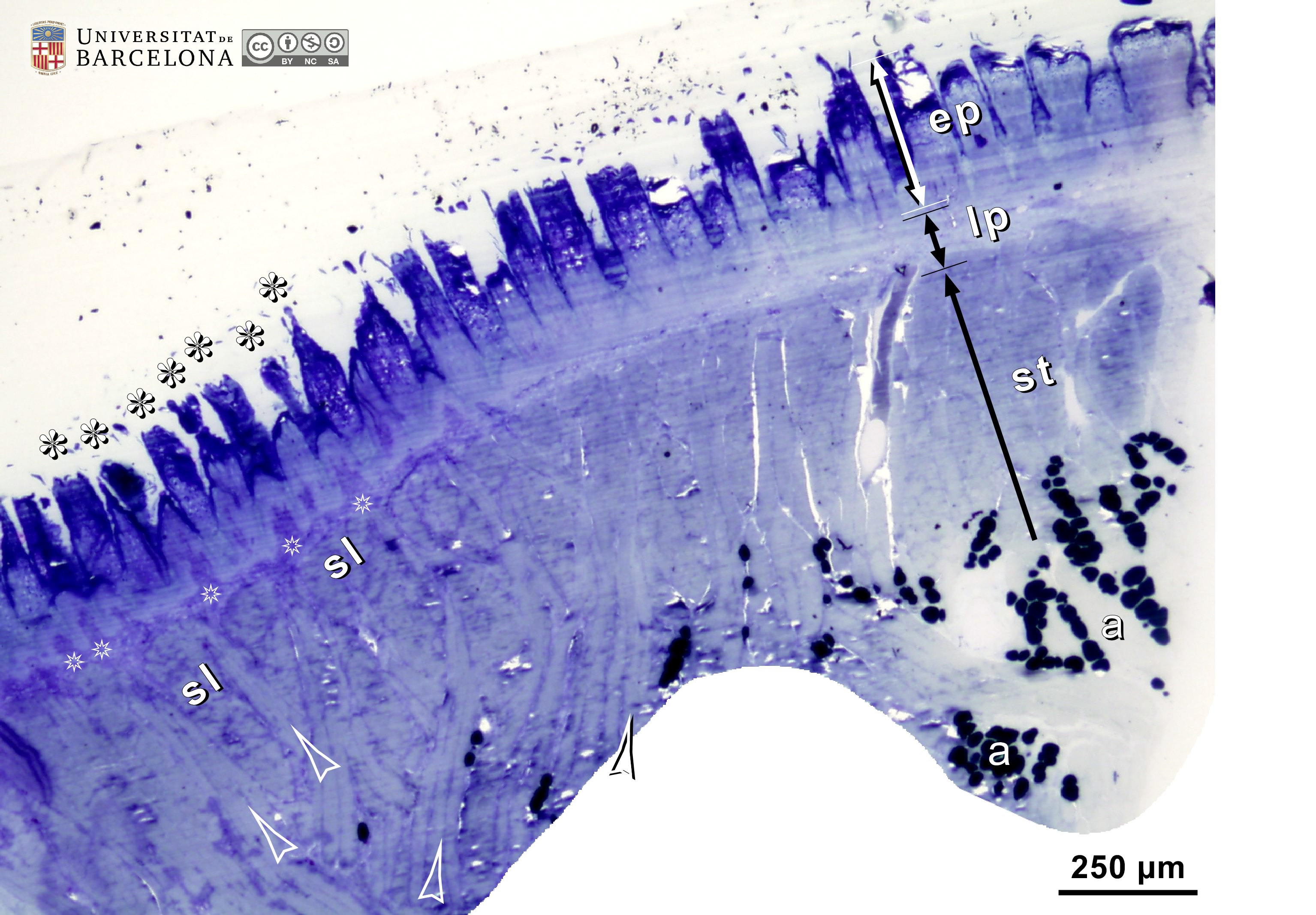 View/Open |
| P_O_tongue_semithin_2um_4x_b087.jpg | Dorsal surface of the rat tongue. No labelling. | 791.03 kB | JPEG |  View/Open |
| P_O_tongue_semithin_2um_20x_b088_b089_collage_LETTERS.jpg | Dorsal surface of the rat tongue (collage of micrographs allowing moderate zoom). The epithelium (ep) is thick, with conical protrusions – the filiform papillae (white asterisks) –. The epithelial stratum granulosum (gra) contains small keratohyaline granules stained blue. The lamina propria is thin and slightly violet, with regular elevations (white stars) that push the epithelium upward, contributing to the formation of papillae. Beneath this layer, thick skeletal muscle fibres are cut transversally as in the superior longitudinal muscles (sl) or longitudinally as in the vertical muscles (ve). In these cases, the striated pattern due to muscle sarcomeres can be seen (empty arrowheads). Osmium tetraoxide postfixation, epoxy section 2 µm thick, toluidine blue stain. | 893.36 kB | JPEG |  View/Open |
| P_O_tongue_semithin_2um_20x_b088_b089_collage.jpg | Dorsal surface of the rat tongue (collage of micrographs allowing moderate zoom). No labelling. | 859.75 kB | JPEG | 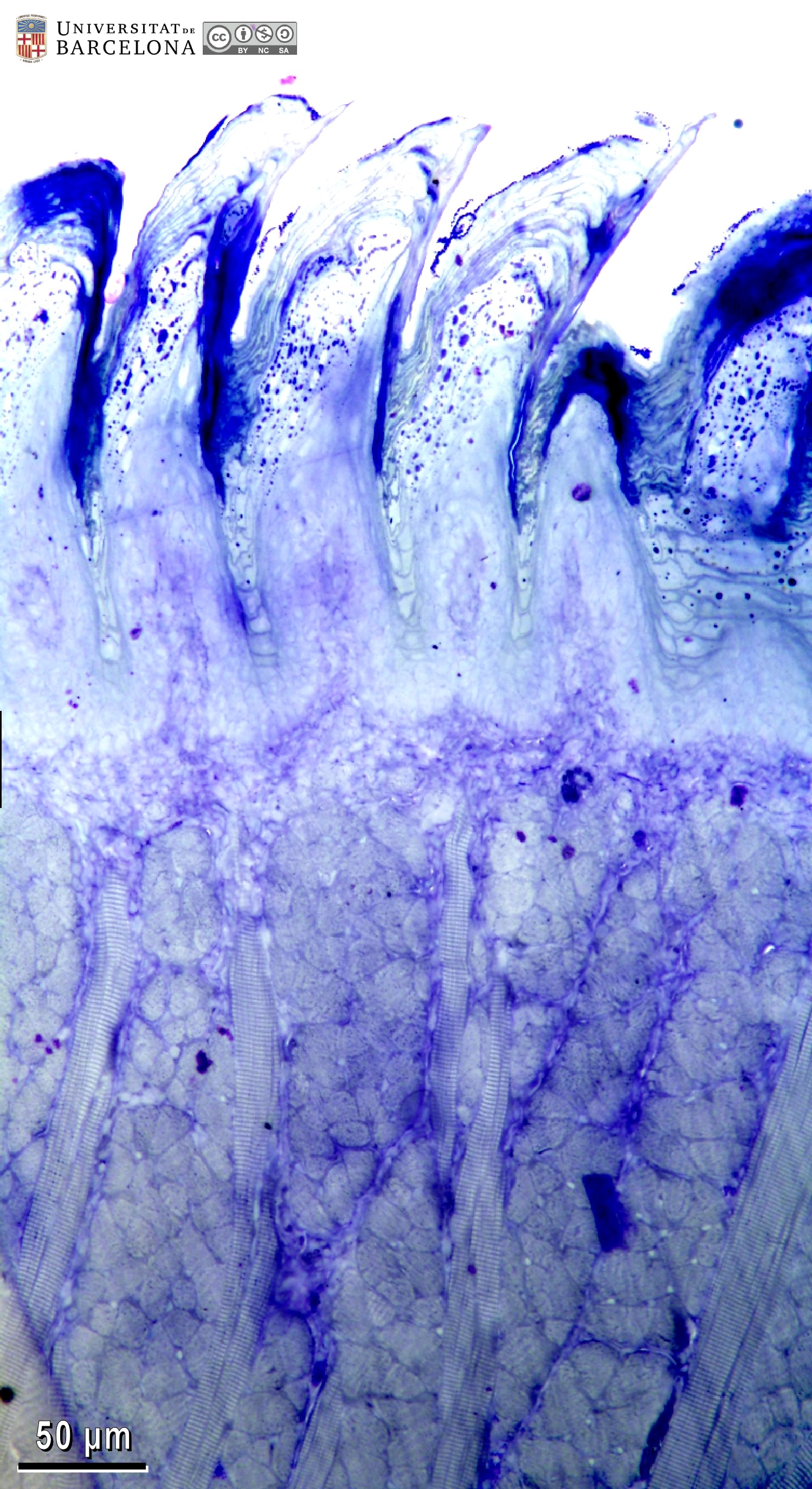 View/Open |
| P_O_tongue_semithin_2um_50x_05_07_collage_LETTERS.jpg | Detail of lingual papillae in the rat tongue (collage of micrographs allowing detailed zoom). In the epithelium, staining in the strata basale (ba) and spinosum (sp) faded away due to slide aging, but cell borders at the stratum granulosum are distinct, and keratohyaline granules can be seen within the cells. Some cell nuclei with light chromatin and a distinct nucleolus (white arrowheads) can be seen when zooming in. The stratum corneum (cor) of dead cells is heavily stained. The connective tissue in the lamina propria (lp) becomes taller at the papillae (empty star) bulging into the epithelium, forming cone-shaped elevations, the filiform papillae (fi). Osmium tetroxide postfixation, epoxy section 2 µm thick, toluidine blue stain. m, muscular fibres. | 1.1 MB | JPEG |  View/Open |
| P_O_tongue_semithin_2um_50x_05_07_collage.jpg | Detail of lingual papillae in the rat tongue (collage of micrographs allowing detailed zoom). No labelling. | 1.04 MB | JPEG | 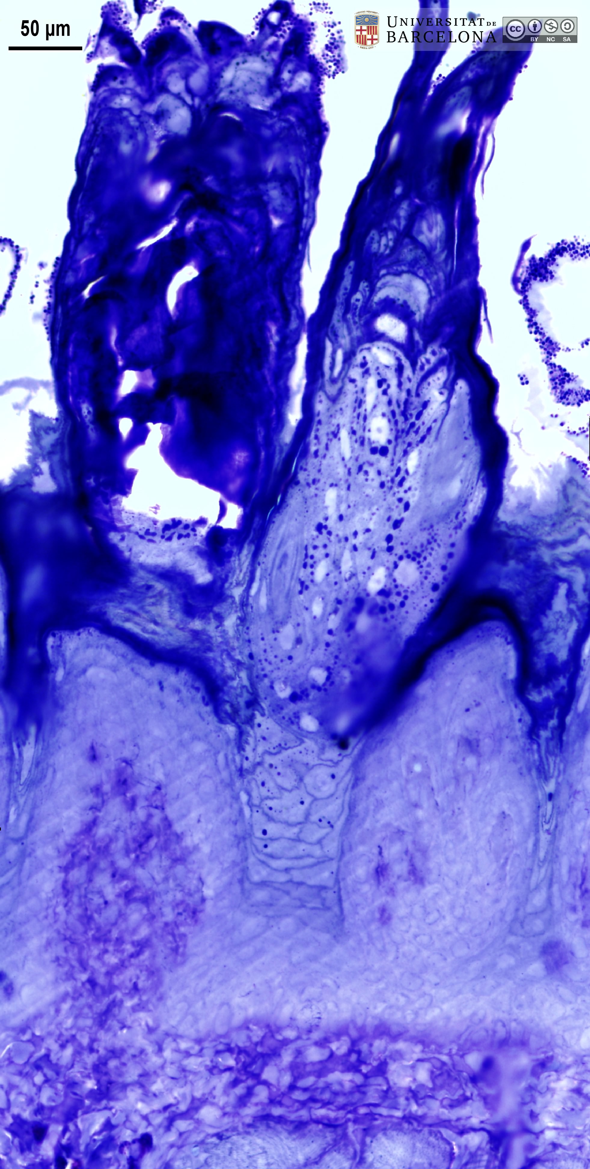 View/Open |
| P_O_tongue_semithin_2um_50x_12_LETTERS.jpg | Connective tissue and skeletal muscle deep within the rat tongue. Two arteries (a) are seen in the connective tissue. Their wall consists of an inner elastic fibre layer (iel) and a ring of smooth muscle cells (sm). Various mast cells with metachromatic granules (stained pink) can be seen. The skeletal (striated) muscle bundles (st) were cut longitudinally and the striated pattern of the myofibrils is visible. Dark spots (empty arrowheads) probably are glycogen deposits. Osmium tetroxide postfixation, epoxy section 2 µm thick, toluidine blue stain. | 1.55 MB | JPEG | 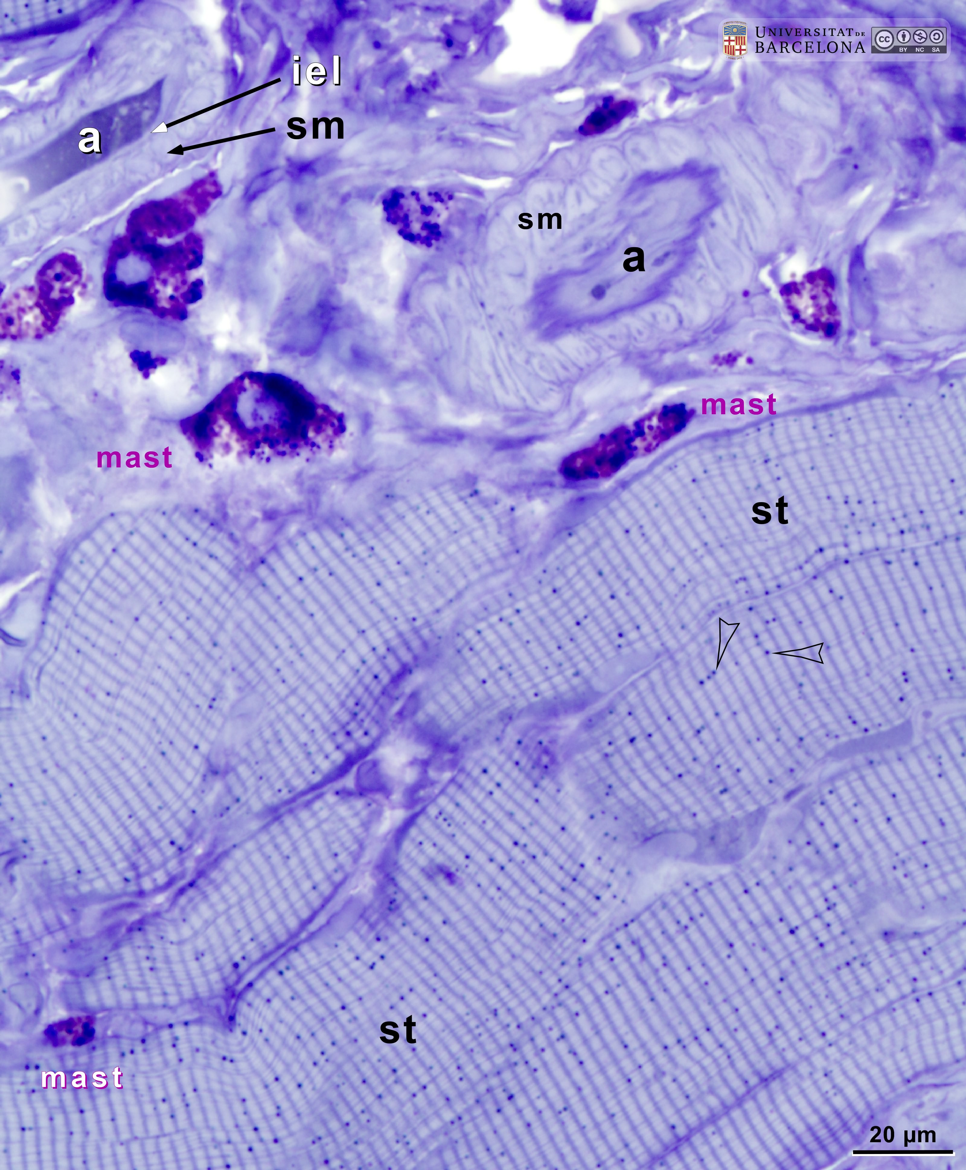 View/Open |
| P_O_tongue_semithin_2um_50x_12.jpg | Connective tissue and skeletal muscle deep within the rat tongue. No labelling. | 1.51 MB | JPEG | 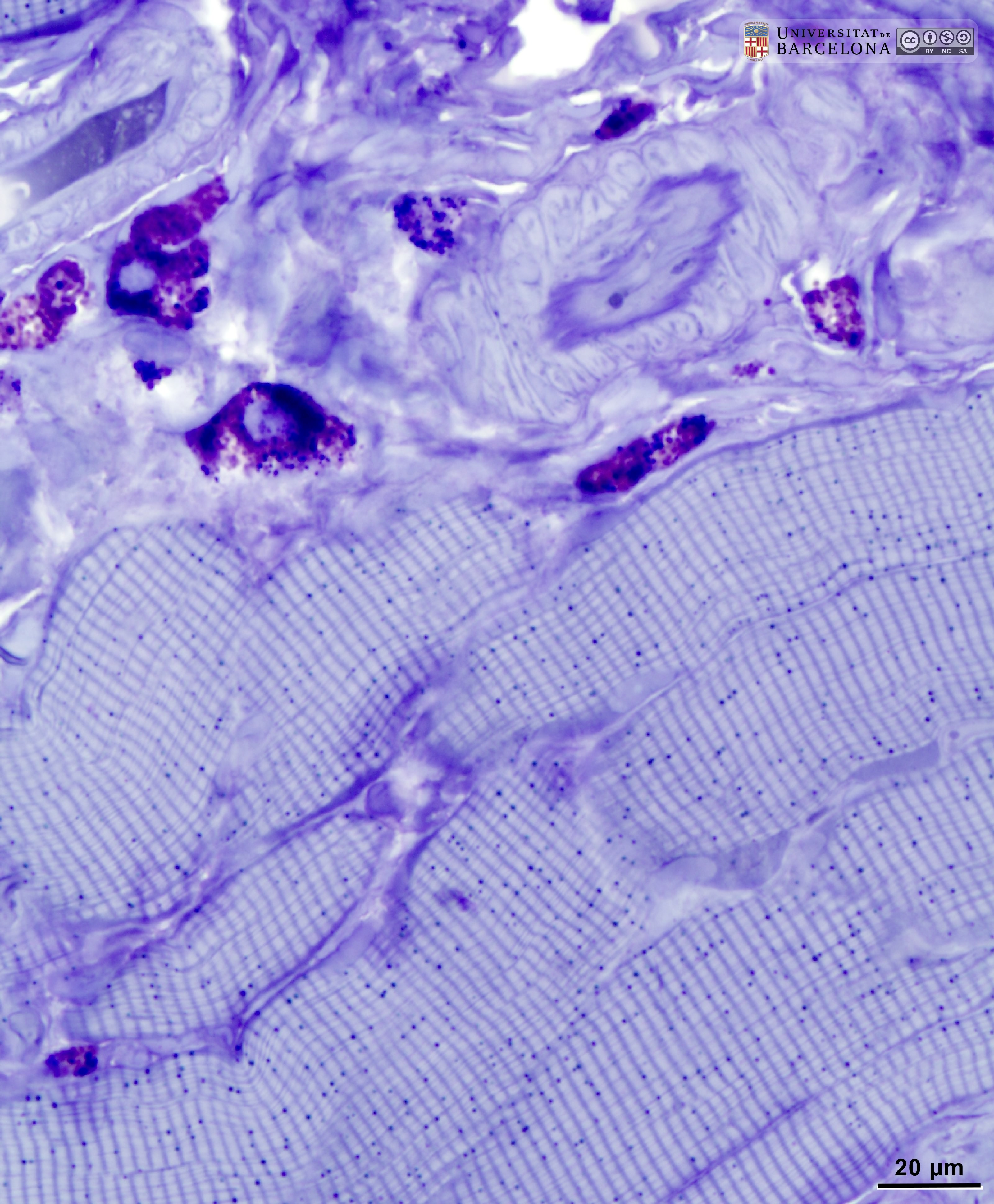 View/Open |
| P_O_tongue_semithin_2um_50x_10_LETTERS.jpg | Skeletal muscles in deep regions of the rat tongue. When muscle fibres are sectioned longitudinally (l), the regular striated pattern of the myofibrils is observed. The small dark dots (empty arrowheads) probably are glycogen deposits. When muscle fibres are cut transversally (t), myofibrils appear as light blue dots, not to be confused with the dark blue deposits (arrowheads). In the connective tissue between the muscle bundles, two mast cells with metachromatic granules can be seen. A small nerve branch (n) with darkly stained myelin sheaths is also seen. Osmium tetroxide postfixation, epoxy section 2 µm thick, toluidine blue stain. | 960.74 kB | JPEG | 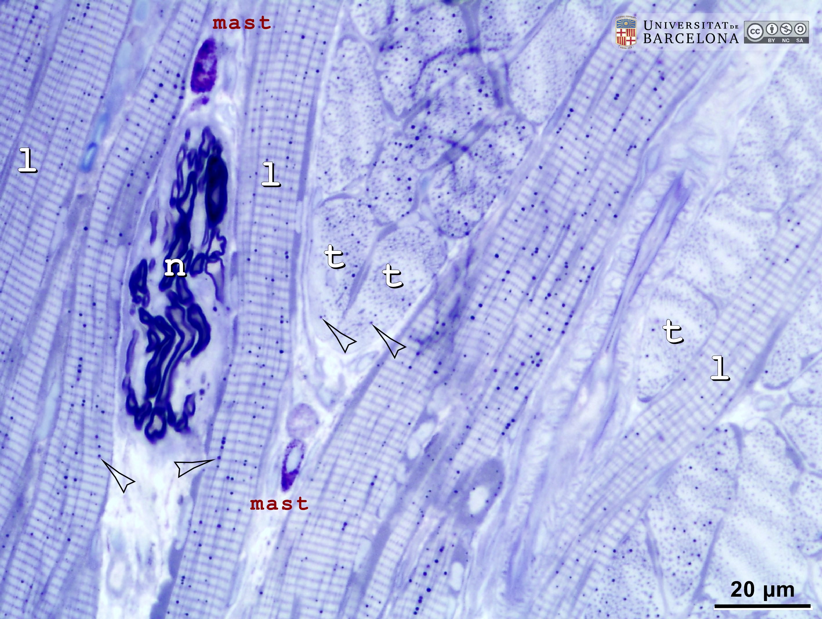 View/Open |
| P_O_tongue_semithin_2um_50x_10.jpg | Skeletal muscles in deep regions of the rat tongue. No labelling. | 939.15 kB | JPEG | 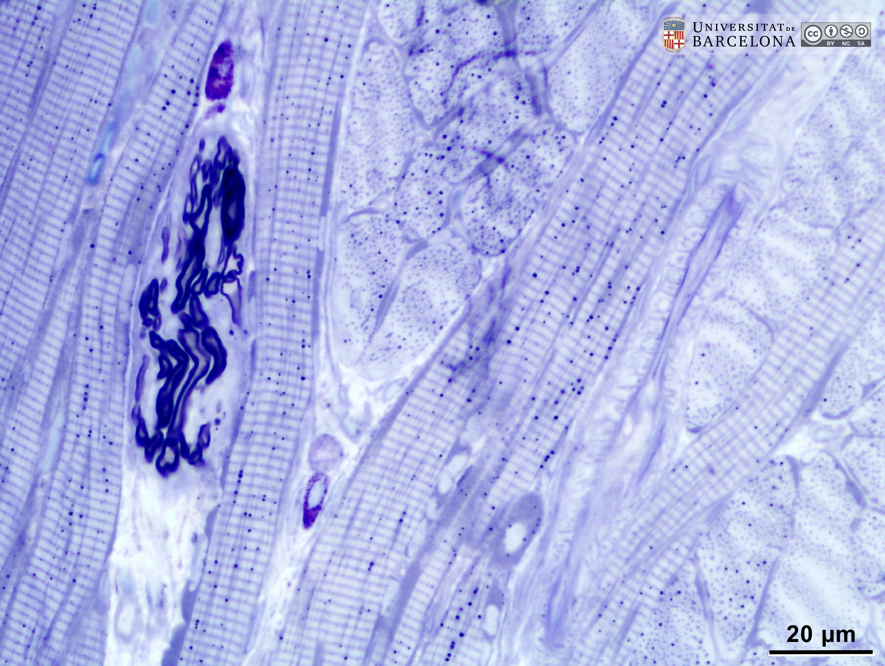 View/Open |
| P_O_tongue_semithin_2um_100x_16_LETTERS.jpg | Skeletal muscle fibres and nerves in the rat tongue. In longitudinal sections, muscle fibres (stˬl) show the regular pattern of alternating light and dark bands or striae. In transverse sections (stˬt), the myofibrils are seen as pale spots (dashed circle). Dark blue dots (empty arrowheads) are seen near the microfibrils, possibly representing glycogen accumulations. Two nerve branches are seen between the muscle fibres. These branches are surrounded by a thin perineurium and contain myelin sheaths (my). Osmium tetroxide postfixation, epoxy section 2 µm thick, toluidine blue stain. | 995.68 kB | JPEG | 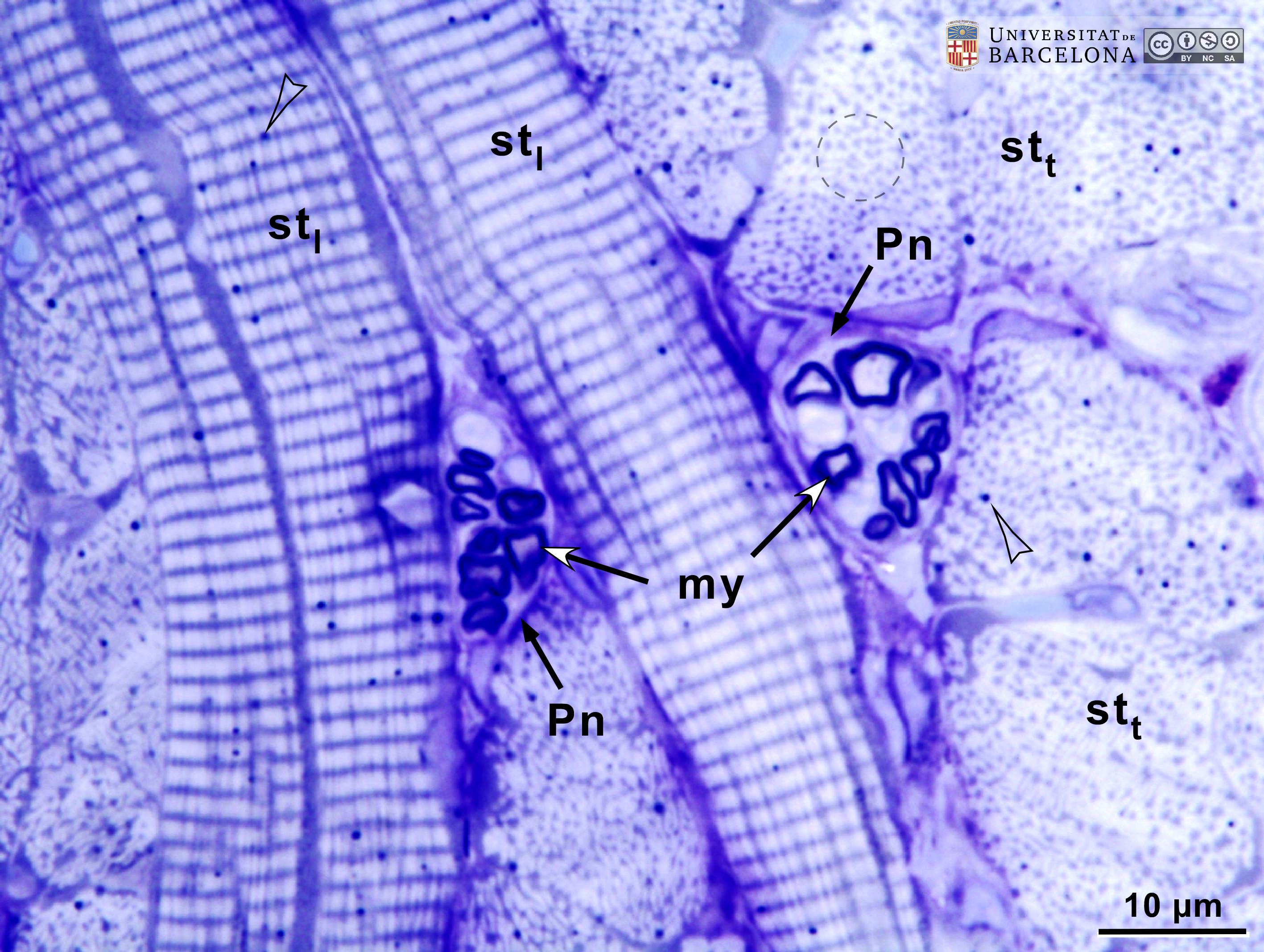 View/Open |
| P_O_tongue_semithin_2um_100x_16.jpg | Skeletal muscle fibres and nerves in the rat tongue. No labelling. | 971.77 kB | JPEG |  View/Open |
| P_O_tongue_semithin_2um_50x_18_LETTERS.jpg | Nerve branches in the rat tongue faintly stained with toluidine blue. The nerve branches (n) are surrounded by a thin perineurium (black arrows) and possibly an epineurium surrounding the central branch. These nerves contain multiple myelin sheaths stained black due to osmium tetroxide postfixation. The lipid drop in white adipocytes (wa) is black as well. The surrounding connective tissue contains fibroblasts (f), lightly stained collagen fibres (col), and blood vessels (v). Epoxy section 2 µm thick, toluidine blue stain. | 673.85 kB | JPEG | 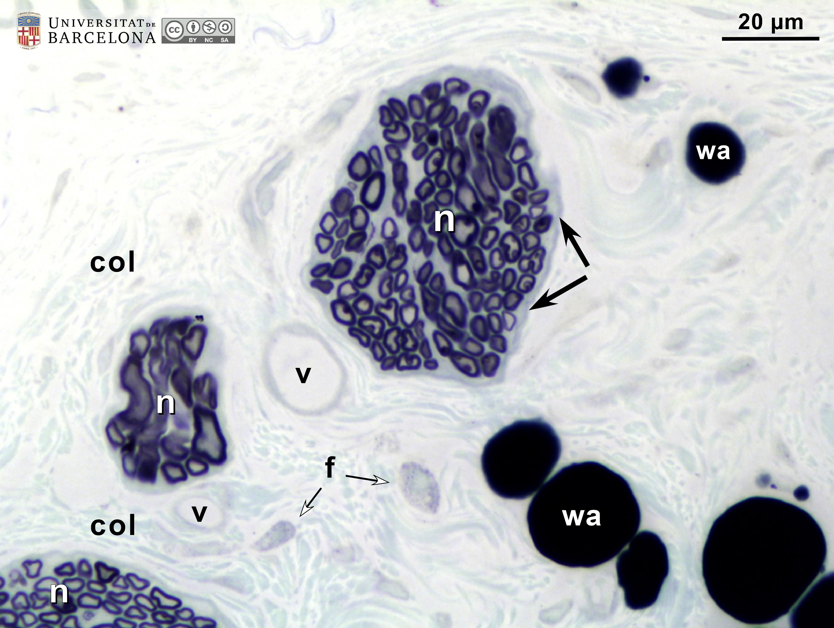 View/Open |
| P_O_tongue_semithin_2um_50x_18.jpg | Nerve branches in the rat tongue faintly stained with toluidine blue. No labelling. | 651.73 kB | JPEG | 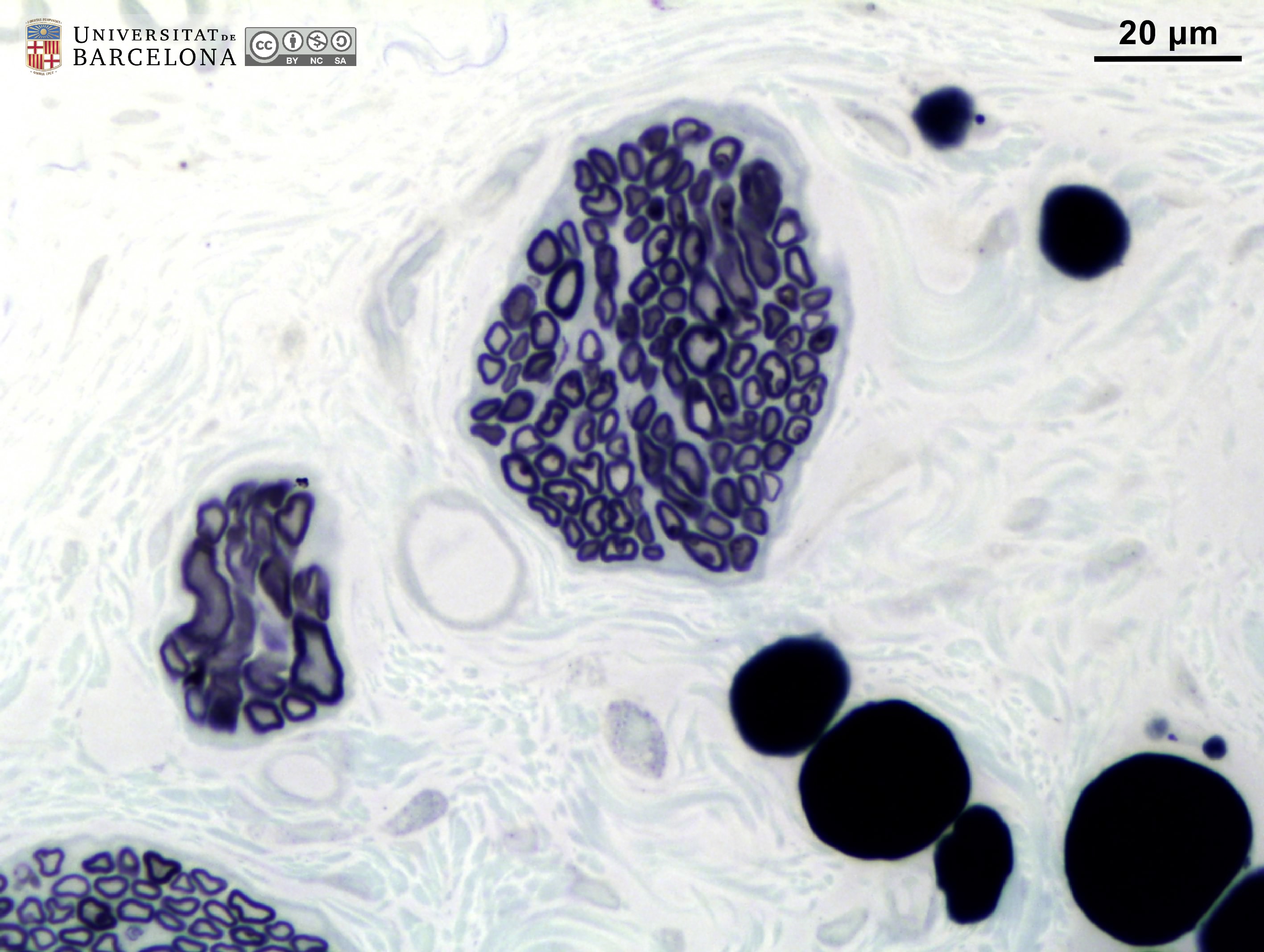 View/Open |
| P_O_tongue_semithin_2um_100x_20_LETTERS.jpg | Nerve in the rat tongue. A perineurium (black arrows) surrounds the nerve, which may also be enclosed by an epineurium (not labelled). The perineurium divides the nerve diagonally (arrowheads), forming two (1 and 2) or possibly three (3) bundles. Within each bundle, axons are surrounded by a myelin sheath (my), which is stained black due to osmium tetroxide postfixation. Epoxy section 2 µm thick, faint toluidine blue staining. col, faintly stained collagen fibres. | 714.25 kB | JPEG | 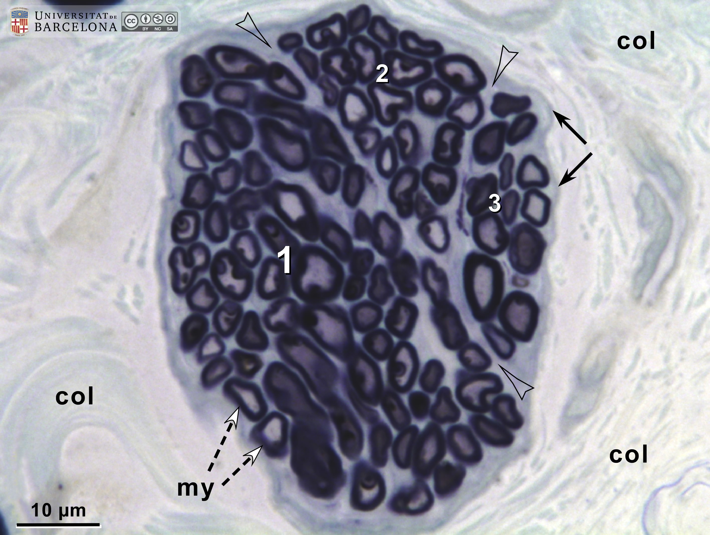 View/Open |
| P_O_tongue_semithin_2um_100x_20.jpg | Nerve in the rat tongue. No labelling. | 684.79 kB | JPEG | 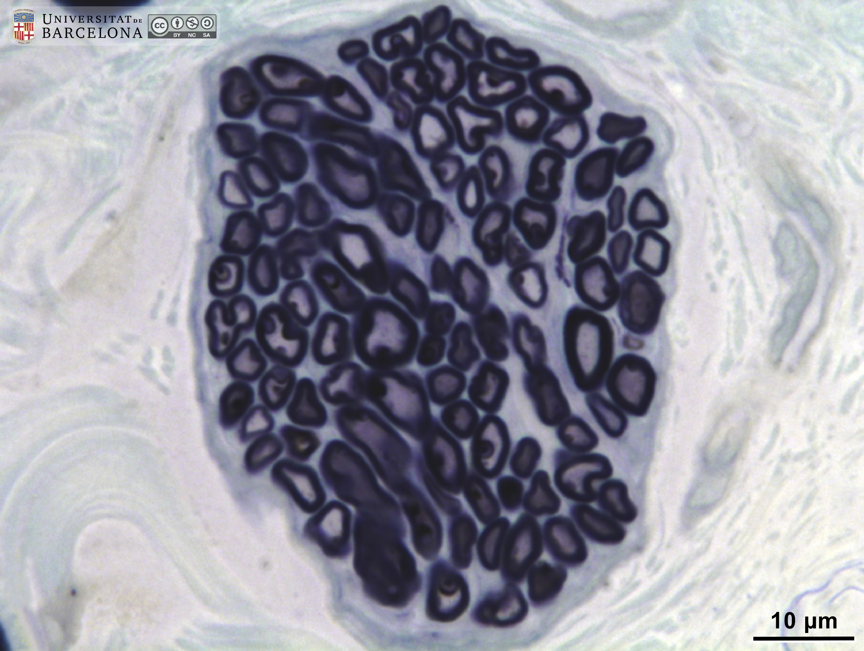 View/Open |
This item is licensed under a
Creative Commons License